


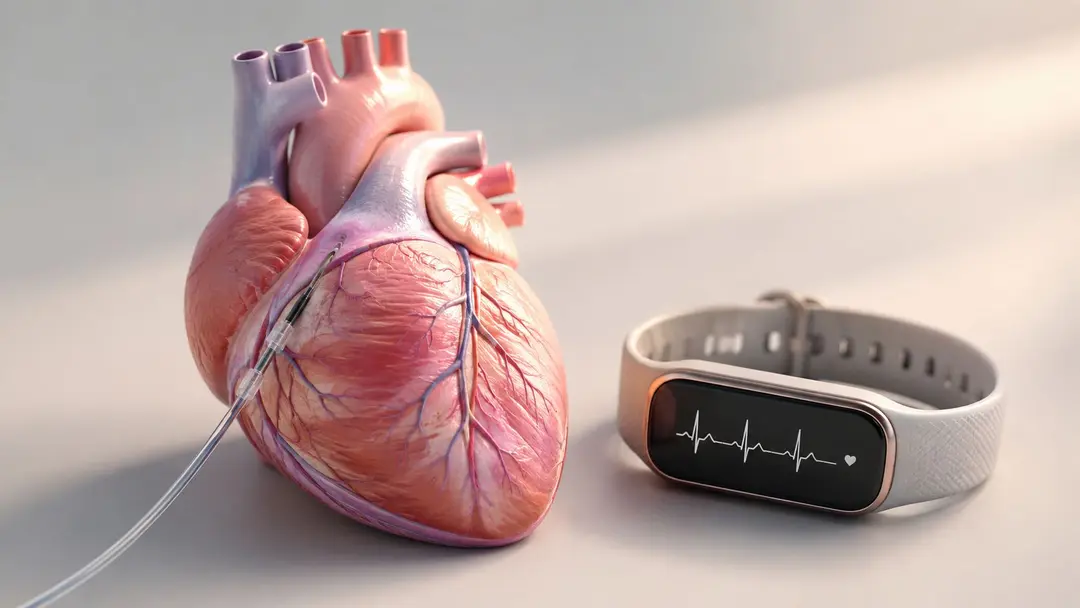


Hello Heart Hero. You're home after a scheduled or recent cardiac catheterization, and every ache, bruise, skipped beat, or unfamiliar sensation may suddenly feel important. That anxiety is understandable. Paying close attention doesn't mean something is wrong, it means you're trying to recover safely.
Searching for cardiac catheterization complications often brings up a frightening list led by stroke or death. Those events matter, but the issues you're more likely to notice involve the wrist or groin used for access, bruising, bleeding, soreness, or changes in the pulse. Some problems are quieter, including radial artery occlusion, which may cause few symptoms and can affect future access.
This guide explains what the procedure involves, how diagnostic and interventional catheterization differ, which complications deserve attention, and which symptoms require urgent help. It also covers practical questions for your care team, recovery habits, and the sensible role of wearable ECG tracking between appointments. Settle in, read at your own pace, and save questions for your next cardiology visit.
Welcome, Heart Hero
Start with the sensations you can feel
Your wrist may feel tender when you move it, the bandage may have left a bruise, and your heart may give an occasional flutter. You might wonder whether to rest, call the clinic, or seek emergency care. The right response depends on the symptom, how intense it is, whether it is worsening, and what your procedure involved.
Mild soreness or bruising around the access site can fit ordinary healing, but keep watching it. Rapid swelling, ongoing bleeding, a cold or pale hand, severe pain, chest pressure, sudden weakness, or major breathing difficulty needs prompt attention.
A calm recovery rule: Notice the change, record what is happening, and use the discharge instructions from your own clinical team as the first point of reference.
The reassuring bigger picture
Diagnostic catheterization has become safer over time. A historical review of cardiac catheterization safety describes mortality falling from about 1% in the early 1960s to about 0.08% by the 1990s, reflecting long-term improvements in technique and care. Contemporary summaries generally describe major complications during diagnostic procedures as uncommon, while recognizing that risk is not zero.
The procedure itself shapes that risk. A diagnostic study examines the heart and coronary arteries. An intervention may include balloon treatment, stent placement, or complex valve work, so its recovery questions can differ. Your age, other illnesses, kidney function, blood-thinning medicines, access route, and clinical stability also matter.
The goal is practical awareness, not alarm. As you continue, you will learn to tell access-site problems from rhythm symptoms, understand why a quiet wrist issue can matter, recognize warning signs that need immediate help, and collect useful details for your clinicians. Wearable ECG tracking may also provide a simple record of rhythm changes during recovery, without replacing medical advice or follow-up care.
What Cardiac Catheterization Actually Is
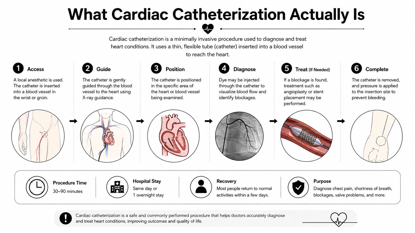
Cardiac catheterization uses a thin, flexible tube, called a catheter, that travels through a blood vessel toward the heart. Clinicians may use it to inspect coronary arteries, measure pressures, inject contrast, place a stent, or support valve and structural-heart procedures.
A simple three-part mental model helps:
- Access: The team enters a blood vessel, often at the wrist through the radial artery or at the groin through the femoral artery.
- Procedure: Catheters, wires, imaging, pressure measurements, and sometimes contrast help the team diagnose or treat a heart problem. Fluoroscopy is the moving X-ray guidance used during many catheter-based procedures. You can learn more about what fluoroscopy is.
- Recovery: The team removes the catheter, controls bleeding, applies a closure device or compression band, and monitors you while the vessel begins to heal.
Diagnostic and interventional procedures are different
A diagnostic cath gathers information. The operator may look for narrowing, evaluate blood flow, measure pressures, or assess valve function. An interventional cath adds treatment, such as angioplasty, stent placement, or another catheter-based repair.
That distinction matters because intervention usually involves more catheter manipulation, larger or longer devices, additional medicines, and more time inside the blood vessels. In one modern cohort, diagnostic procedures had a lower complication burden than therapeutic procedures in earlier mixed-practice data, where overall complications were 8%, diagnostic procedures were 3.6%, and therapeutic procedures were 15.1% (large left-heart catheterization cohort and complication analysis).
Why the access route changes the risk
Radial access enters through the wrist and often makes bleeding easier to control because the artery is close to the skin and can be compressed. Femoral access enters through the groin and may be preferred when the equipment, anatomy, or procedure requires it.
Neither route is automatically right for every person. Vessel size, previous procedures, circulation in the hand or leg, urgency, operator experience, and planned equipment all influence the choice. Categorizing a complication by where it begins and when it appears helps make unfamiliar clinical language easier to understand.
The Major Complication Types You Should Know
The most useful way to think about risk is not as one frightening percentage. It's as a group of possible problems with different warning signs and different levels of urgency.
A large modern cohort of 43,786 diagnostic left-heart catheterizations reported 36 major complications, an incidence of 0.082%, or 8.2 per 10,000 procedures (modern left-heart catheterization study). Summarized reviews place diagnostic mortality around 0.05% and major complications generally below 1% (NCBI overview of cardiac catheterization). Those figures apply to specific populations and procedures, not automatically to every cath.
A practical comparison
| Complication Type | Approximate Incidence | Key Risk Factors |
|---|---|---|
| Access-site bleeding, hematoma, pseudoaneurysm, or vessel injury | Vascular complications occurred in 1.0% overall in one 7,690-procedure series, with surgical repair required in 0.5% (vascular complication series) | Older age, female sex, larger sheaths, longer procedures, anticoagulant or antiplatelet treatment, and interventional procedures |
| Major complications after diagnostic left-heart catheterization | 0.082%, or 8.2 per 10,000 in a cohort of 43,786 procedures (modern left-heart catheterization study) | Comorbidity, unstable illness, vascular anatomy, and procedure complexity |
| Stroke | Around 0.05% to 0.1% for diagnostic cases in contemporary summaries (historical and contemporary safety review) | Procedure type, atherosclerosis, age, instability, and catheter manipulation |
| Death during diagnostic procedures | Around 0.05% in contemporary summaries (NCBI overview of cardiac catheterization) | Severe illness, shock, arrhythmia, infection, and procedural complexity |
| Radial artery occlusion | 3.0% with standard hemostasis and 0.9% with prophylactic ipsilateral ulnar compression at 30 days in a 2025 trial of 3,000 diagnostic patients (SCAI radial artery occlusion guidance) | Compression technique, sheath and vessel size, anticoagulation timing, and loss of blood flow during hemostasis |
| Vascular complications by access route | Femoral 1.3%, radial 0.0%, and brachial 4.5% in one 1998 to 2007 study (access-route comparison) | Access anatomy, procedure type, device size, and operator technique |
These categories overlap. A hematoma is blood collecting under the skin. A pseudoaneurysm is a leak that forms a contained pocket beside the artery. An arteriovenous fistula is an abnormal connection between an artery and vein. A dissection means the vessel wall has been injured and blood has entered between its layers.
Other complications can affect the heart, kidneys, lungs, or immune system. Temporary arrhythmias may occur during catheter manipulation. Heart attack, stroke, severe allergic reaction, contrast-associated kidney injury, infection, and cardiac tamponade are serious possibilities, but their likelihood varies sharply by procedure and patient condition.
If you take clopidogrel or another antiplatelet drug, don't stop it on your own after a stent. A plain-language review of clopidogrel side effects can help you prepare questions, but your prescribing clinician must decide whether any medication should change.
The Quiet Complication Most Guides Miss
The most dramatic complication isn't always the most practically important one. Radial artery occlusion, or RAO, can develop after wrist access while causing little or no discomfort. Because many people still have blood reaching the hand through another artery, the hand may look and feel normal even when the radial artery is no longer open.
Specialty guidance warns that RAO can be underestimated because many patients are asymptomatic (SCAI guidance on reducing radial artery occlusion). That quietness matters. An occluded radial artery may limit future wrist access, and it can become more relevant if another catheter procedure is needed later.
What raises the risk
The access device can press against the vessel, and compression used to stop bleeding can temporarily reduce blood flow. Risk may rise when the sheath is relatively large for the artery, anticoagulation is not timed appropriately, or hemostasis stops flow entirely instead of allowing blood to continue moving through the vessel.
The same 2025 trial found that 30-day RAO fell from 3.0% with standard hemostasis to 0.9% with prophylactic ipsilateral ulnar compression in 3,000 diagnostic catheterization patients (SCAI trial summary). That result illustrates why the details of wrist-band management and follow-up matter.
Access problems aren't limited to the wrist. In a 7,690-procedure series, vascular complications included bleeding, hematoma, pseudoaneurysm, arteriovenous fistula, thromboembolism, and infection. The overall rate was 1.0%, with surgical repair required in 0.5%, and the risk was higher after coronary angioplasty than angiography alone, 3% versus 1% (vascular complication series).
Before leaving, ask:
- Access choice: Why is radial or femoral access better for my anatomy and procedure?
- Patency check: Will someone check that blood is still flowing through the radial artery?
- Compression plan: How long should the band or dressing remain in place?
- Follow-up: Which changes in pulse, color, temperature, or sensation should prompt a call?
Get your ECG checked by certified experts within minutes on the Qaly app.


Red Flags to Watch For After the Procedure
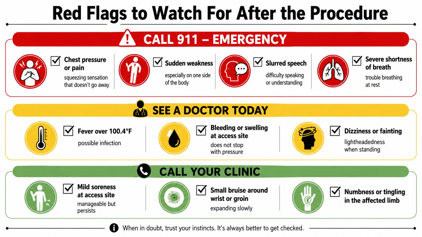
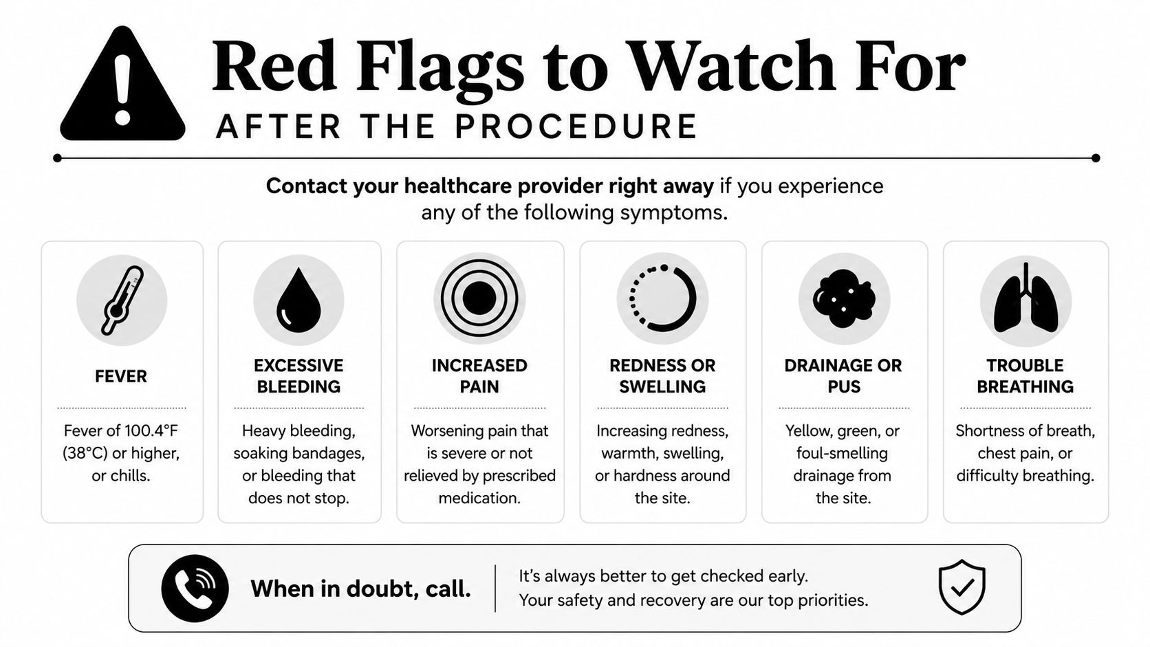
Discharge instructions from your own team take priority, but urgency can be organized by symptom severity and timing. The first hours are especially important for active bleeding, sudden chest symptoms, breathing difficulty, neurological changes, and rapidly expanding swelling.
Call emergency services immediately
Call 911 or your local emergency number for chest pressure that doesn't go away, severe shortness of breath at rest, sudden weakness, facial drooping, slurred speech, confusion, or fainting with ongoing symptoms. These signs can indicate a heart or neurological emergency, and you shouldn't drive yourself.
Rapid swelling at the wrist or groin, bleeding that doesn't stop with firm direct pressure, or a limb that becomes cold, pale, blue, severely painful, or suddenly weak also needs emergency assessment. Don't remove a compression device unless your discharge instructions tell you to do so.
Contact the clinical team the same day
Call your clinic or the after-hours cardiology number for an expanding bruise, persistent oozing, increasing access-site pain, new numbness or tingling, or a change in hand or foot color. Fever can signal infection, particularly when it occurs with redness, warmth, drainage, or worsening tenderness.
Pink or red urine after femoral access deserves prompt advice because bleeding behind the abdominal lining can be difficult to see externally. Lightheadedness when standing, unusual weakness, or repeated palpitations also shouldn't be dismissed, especially if these symptoms feel different from what you experienced before the procedure.
For a plain-language symptom refresher, keep cardiac event symptoms to recognize available with your discharge paperwork.
Record before you call
Write down when the symptom began, whether it's worsening, your pulse if you can measure it safely, any visible change at the access site, and the medicines you've taken. If a wearable records an unusual rhythm, save the tracing rather than repeatedly checking it while becoming more anxious.
Some issues appear later. Delayed bleeding may follow a dose of an anticoagulant or antiplatelet medicine, and a contrast-related reaction can occur after you've left the facility. New rash, swelling, wheezing, reduced urination, or worsening breathlessness warrants medical guidance rather than guesswork.
Tracking Your Heart Rhythm at Home After a Cath
A wearable ECG can add useful information when recovery includes a new flutter, racing sensation, skipped beat, or unexplained lightheadedness. Most consumer devices record a single-lead tracing, which can help identify patterns such as atrial fibrillation, a slow rate, pauses, or frequent premature beats. It isn't a replacement for the clinical assessment, a 12-lead ECG, blood tests, imaging, or a multi-day Holter or patch monitor.
Use the device to answer a question
Take a recording when symptoms occur, if you can do so without delaying emergency care. A second reading after you've rested may show whether the sensation persists, but repeated recordings can also increase anxiety without adding meaningful information.
Keep a simple log:
- Symptom: What did you feel, such as fluttering, pounding, pause, dizziness, or breathlessness?
- Timing: When did it start, and how long did it last?
- Context: Were you resting, walking, dehydrated, upset, or taking a new medicine?
- Tracing: Save the ECG and label it with the symptom and time.
- Response: Note whether rest helped and whether the symptom returned.
Trends can be more useful than one unusual-looking snapshot. Your cardiology team may care about repeated rhythm patterns, changes in resting rate, or shifts in measured PR and QTc intervals. A wearable's interval estimate can be affected by signal quality, heart rate, movement, and the device itself, so an apparent change needs clinical interpretation.
Important boundary: A wearable can document a rhythm, but it can't reliably rule out a blocked artery, internal bleeding, pulmonary problem, or other emergency.
Human review may help you organize information between visits. Qaly connects wearable ECG recordings with certified cardiographic technicians for rhythm interpretation, which can give you a clearer record to discuss with your clinician. If a tracing looks strange alongside chest pressure, fainting, severe breathlessness, or neurological symptoms, seek urgent care rather than waiting for an app interpretation.
For background on the role of home recordings, see this guide to heart rhythm monitoring. The goal is not to chase every irregular line. It's to capture meaningful episodes, describe symptoms accurately, and know when technology should give way to direct medical care.
Prevention, Consent, and Recovery That Works
Good preparation won't eliminate every complication, because some risks depend on vessel anatomy, procedure complexity, and the clinical team. It can reduce avoidable confusion and help clinicians identify risks before the catheter enters the vessel.
Before the procedure
Bring a complete medication list, including aspirin, clopidogrel, anticoagulants, diabetes medicines, supplements, and any previous contrast reaction. Ask whether kidney function needs checking, what hydration plan is appropriate, and which medicines should be taken or held. Never stop an antiplatelet or anticoagulant without explicit instructions, particularly after stent placement.
Ask direct consent questions:
- Purpose: Is this diagnostic, interventional, or potentially both?
- Access: Which route is planned, and why does it fit my anatomy?
- Bleeding: How does my medication history affect bleeding risk?
- Operator and team: Who will perform the procedure, and who should I contact afterward?
- Recovery: How will you check the access site and, when relevant, radial artery flow?
Clear consent should leave you understanding the expected benefit, meaningful alternatives, and what would make the team change the plan. If your facility uses digital documentation, resources such as Closer Innovation Labs Corp. healthcare can help explain how electronic patient-consent workflows are organized, though your clinical team remains responsible for the medical discussion.
After you return home
Follow the written activity and wound-care instructions. Keep the access site visible enough to inspect, avoid changing medicines independently, pace activity, and attend scheduled follow-up. Combine symptom notes with wearable ECG recordings only when rhythm symptoms are part of your recovery concern.
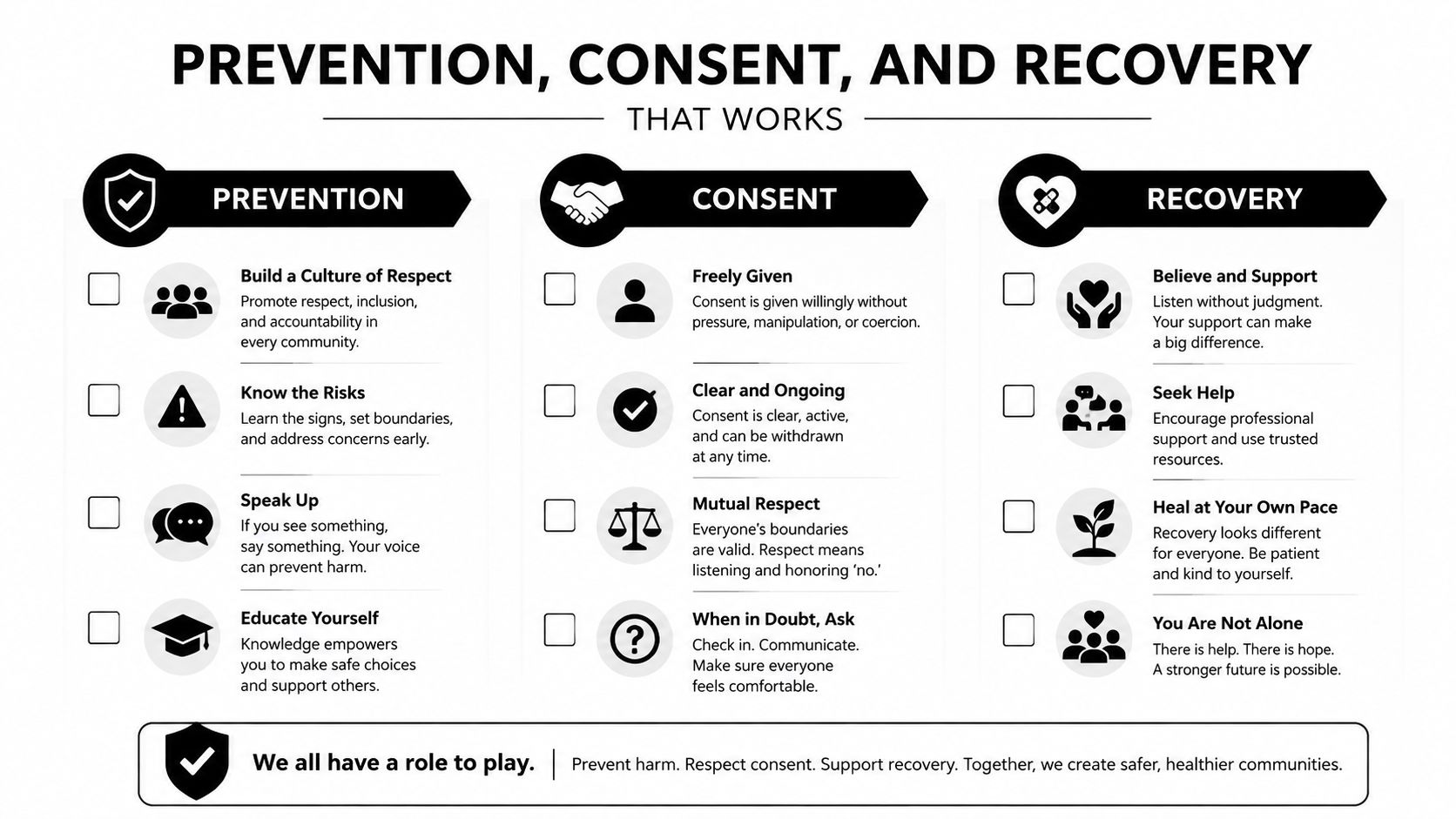
A screenshot-friendly checklist
- Medication review: Confirm exactly what to take, hold, or restart.
- Kidney plan: Ask whether kidney function and hydration need attention.
- Access plan: Understand why the wrist or groin route was selected.
- Bleeding plan: Know how to respond to bleeding and when pressure isn't enough.
- Pulse and limb check: Ask what changes in temperature, color, sensation, or pulse matter.
- Rhythm record: Save symptom-linked ECGs, but don't delay emergency care.
- Follow-up route: Keep the clinic and after-hours numbers where family can find them.
What Most Heart Heroes Want to Know Next
How long will bruising last? Bruising and soreness often improve gradually, but an expanding bruise, increasing pain, persistent bleeding, or new numbness deserves a call.
Is chest discomfort normal? Mild tenderness can occur, but pressure, squeezing, breathlessness, sweating, fainting, or pain that feels like your original heart symptoms should be treated urgently. Don't try to diagnose it from home.
Is radial access safer than femoral access? The risk profile differs. One study reported fewer vascular complications with radial access than femoral access, while brachial access had a higher rate in that dataset (access-route comparison). Your anatomy and procedure still matter.
When can I drive or exercise? Follow your discharge instructions, because timing depends on access site, treatment, symptoms, and the clinician's assessment.
What if my wearable ECG looks strange? Save the tracing and contact your care team for interpretation. If it comes with severe symptoms, call emergency services first.
Clear communication improves recovery. These patient experience tips from CallZent may help you prepare questions and organize follow-up conversations. Complications are uncommon, the most frequent problems are usually manageable, and careful home observation turns uncertainty into useful information.
If you're recovering from cardiac catheterization, save this guide, review your discharge instructions, and write down three questions for your next appointment: Which access-site changes should I report, will my radial artery flow be checked when appropriate, and what rhythm symptoms should trigger urgent care? For wearable ECG users who want human-reviewed rhythm information between cardiology visits, explore Qaly's ECG interpretation service and bring any relevant reports to your clinician.
Noticing rhythm changes after a cath? Certified experts review your ECG within minutes.


