


Key Takeaways
- Your heart beats by creating an electrical signal, which spreads through your heart and squeezes it, pumping blood in and out of your heart with every heartbeat.
- Your ECG's PQRST waves capture this electrical cycle, which produces your each and every heartbeat.
- Your PR, QRS, and QTc Intervals can be powerful clues to the current state of your heart health.
User Profile
Got other questions on your PQRST Intervals? See the complete set of Qaly guides on PQRST Intervals:
- What P Waves Look Like on Your ECG
- QRS Interval on Your ECG - Narrow, Normal, and Wide
- PR Interval on Your ECG - Short, Normal, and Prolonged
- QTc Interval on Your ECG - Short, Normal, and Prolonged
- How to Read an ECG: Stanford Cardiologist Explains
- The Ultimate Cardiologist's Guide to the Smartwatch ECG
Introduction
Hello, heart hero. In your quest to identify that irregular heart rhythm you just felt, you may have come across the terms P Wave, QRS Complex, T Wave, PR Interval, QRS Interval, or QTc Interval. With your trusty watch ECG now in hand, you may be wondering, "What does PR Interval mean on my watch ECG?" Or you might be thinking, "What is a QRS Complex, and is it dangerous if it's wide?" In this guide, we'll help you understand what these terms mean, and what they might indicate about your heart health. Let's dive in.
What's an Electrocardiogram?
Before diving into PR, QRS, and QTc Intervals, it's important to understand exactly what an electrocardiogram, or ECG is. (If you're confident in your understanding of an ECG and what it captures about your heart, though, skip on ahead to the next section for some visual examples of PR, QRS, and QTc Intervals).
To start, remember how your heart beats? It produces an electrical signal, which squeezes and unsqueezes your heart, which in turn pumps your blood to your lungs for oxygen and then out to the rest of your body.
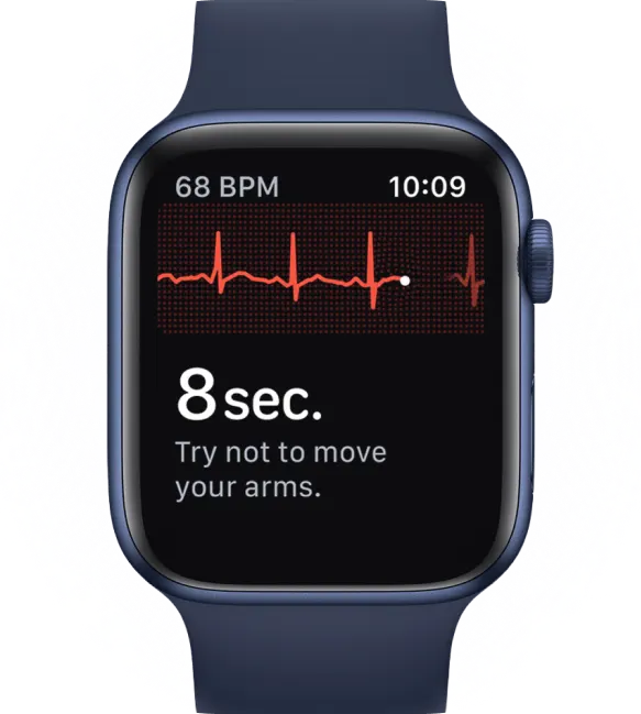
As that electrical signal flows through your heart, your ECG on your watch sees it, and shows it to you as those awesome-looking waves you know as an ECG. Every time your heart completes one heartbeat, it completes one full cycle of that electrical signal flowing through your heart. And as that electrical signal flows through your heart, it produces different waves on your ECG that capture the squeezing and relaxing of your heart with each heartbeat, represented on your ECG as PQRST waves.
So What's PQRST on an ECG?
P Wave
The first wave in the PQRST cycle of each heartbeat is the mighty P Wave. Once your heart's electrical signal starts at the beginning of a heartbeat, it causes your heart's upper chambers, or atria, to squeeze. This squeezes the blood coming into your heart's atria down into your heart's lower chambers, or ventricles. The P Wave on your ECG captures this first squeeze in your heartbeats.

QRS Complex
After the P Wave comes the QRS Complex. You'll recognize this one, since it's usually the tall spike in your ECG. When your heart's electrical signal moves down into your heart's lower chambers, or ventricles, it first moves through specialized wires in your heart called right and left bundle branches (also known as your His-Purkinjee system). These wires help spread your heart's electrical signal as evenly as possible. Your QRS Complex captures your electrical signal spreading down into and through your ventricles, and the beginning of the final squeeze that pushes the blood in your heart back out into the rest of your body.

T Wave
Finally, the T Wave. On your ECG, this third and final wave shows you the unsqueezing, or relaxation of your heart's lower chambers, or ventricles. When your T Wave ends on your ECG, the cycle of blood entering and leaving your heart is complete, and you've completed one full heartbeat.


So What Are PQRST intervals?
Your ECG's PQRST Intervals are measured in seconds, or milliseconds, because that's how "long" or "short" it takes for your heart to beat.
PR Interval
Your PR Interval is measured from the beginning of your P Wave to the beginning of your QRS Complex. It measures how long it takes for your heart's electrical signal to get from the top of your heart to the bottom of your heart.

What’s the Normal Range for My PR Interval?
Your PR Interval is considered normal between 120 milliseconds and 200 milliseconds.
What Does it Mean When My PR Interval Is Prolonged?
Your PR Interval is prolonged at 200 milliseconds or higher. Your PR Interval prolongs when your heart's electrical signal takes a long time to get through your heart's atrioventricular node, or AV node. Your AV node is the “gatekeeper” that sends an electrical signal from the top of your heart to the bottom of your heart. You might see a prolonged PR Interval during sleep, if you exercise frequently, or if you're taking certain medications like beta blockers. You might also see a prolonged PR Interval in your ECG if your AV node isn't functioning properly, causing your heart's electrical signal slows down. For example, here's what first-degree AV block looks like on your watch ECG.
What Does it Mean When My PR interval Is Short?
Your PR Interval is short at 120 milliseconds or lower. Your PR Interval shortens when your heart's electrical signal takes less time than normal to get through your heart's AV node. This can be due to a normal, healthy variation called “enhanced AV node conduction.” You might also see a short PR Interval in your ECG if there's an extra connection between the top and bottom of your heart, called Wolff-Parkinson-White Syndrome. Here's what Wolff-Parkinson-White Syndrome looks like on your watch ECG.
QRS Interval
Your QRS Interval starts at the beginning of your QRS Complex, and ends at the end of your QRS Complex. It measures how long it takes for your heart's electrical signal to spread through your heart's lower chambers, or ventricles.

What’s the Normal Range for My QRS Interval?
Your QRS Interval is considered normal between 80 millioseconds and 120 milliseconds.
What Does it Mean When My QRS Interval Is Wide?
Your QRS Interval is considered wide at 120 milliseconds or higher. Your QRS Interval widens when your heart's electrical signal takes longer than normal to spread through your ventricles. You'll often see this when abnormalities in your heart affect one of those specialized electrical fibers we discussed, called your right and left bundle branches. Scarring in your heart's ventricles, can also cause your heart's electrical signal to spread more slowly through your ventricles, which would lead to a wider QRS Interval. Here's what a wide QRS Interval looks like on your watch ECG.
What Does it Mean When My QRS Interval Is Narrow?
Your QRS Interval is considered narrow at 80 milliseconds or lower. Your QRS Interval narrows when the time it takes for your heart's lower chambers, or ventricles to squeeze becomes shorter. This can happen due to various reasons, including a higher heart rate, changes in how your heart's electrical signal moves through your ventricles, certain medications, and in cases where a bundle branch block (either right or left) is present and then resolves back to normal. It's important to note that unlike wide QRS, a narrow QRS Interval is generally considered normal. Here's what a narrow QRS Interval looks like on your watch ECG.
QTc Interval
Your QT interval starts at the beginning of your QRS Complex and ends at the end of your T wave. The "c" at the end of "QTc" stands for "corrected," and the difference between QT and QTc is that QTc is your QT Interval after it's been "corrected" for heart rate. The reason your QT Interval needs to be corrected is that your QT Interval changes based on your heart rate, so to get a good sense of what's going on with your heart, it helps to correct your QT Interval measurement across your ECG with the QTc Interval. Your QTc Interval represents the time it takes for your heart's ventricles to squeeze the blood out to the rest of your body, and finally, relax.

What’s the Normal Range for My QTc Interval?
Your QTc Interval is considered normal between 350 milliseconds and 450 milliseconds for men, and between 350 milliseconds and 470 milliseconds for women.
What Does it Mean When My QTc Interval Is Prolonged?
Your QTc Interval is considered prolonged at 450 milliseconds or higher for men, and at 470 milliseconds or higher for women. Your QTc Interval prolongs when your ventricle's electrical system isn't working properly. You'll typically see this as a side effect of some medications; however, you might also see this if you have other heart problems like heart failure or electrolyte imbalances. In very rare cases, a genetic abnormality can lead to prolonged QTc Interval. If you're curious, here's what a prolonged QTc Interval looks like on your watch ECG.
What Does it Mean When My QTc Interval Is Short?
Your QTc Interval is considered short at 350 milliseconds or lower. Your QTc Interval can shorten when the your heart's electrical system is working abnormally quickly. The main cause of this is a very rare genetic disorder. Here's what a short QTc Interval looks like on your watch ECG.
Conclusion
Well, that just about wraps up our guide on what your PR, QRS, and QTc Intervals look like on your watch ECG. We hope this could be of some help to you.
If you still need help measuring your ECGs' PQRST Intervals, don't worry, we understand how scary and confusing it can be to experience irregular heartbeats. That's why we created the Qaly app for you and for the hundreds of millions of people around the world who live with heart palpitations and abnormal heart rhythms. On the Qaly app, human experts will measure your ECGs' PR, QRS, and QTc Intervals within minutes for clarity and peace of mind.
To get started with the Qaly app, grab the Qaly app from the App Store or Play Store today. If you have any more questions, or if you need our help in any other way, don't hesitate to reach out to us at support@qaly.co.
As always from the team at Qaly, stay heart healthy ❤️
Have trouble measuring your PR, QRS, or QTc Intervals? On the Qaly app, human experts will measure your ECGs' PR, QRS, and QTc Intervals within minutes. Get started today.


.png)
.png)