





Hello Heart Hero. You're home after a heart attack, the discharge papers are on the kitchen counter, and every unfamiliar sensation feels loaded with meaning. Is the tiredness part of healing? Is that flutter a harmless skipped beat, or a rhythm problem? Should you call your cardiologist, or should you call emergency services?
Those questions are reasonable. Post MI complications aren't all alike, and they don't all require the same response. Some are best handled through scheduled follow-up, while others need emergency care even if the symptom seems brief. This guide focuses on practical decisions, timing, warning signs, testing, and what you can safely track at home. For a broader recovery roadmap, you can also review this heart attack recovery timeline.
A Few Weeks After a Heart Attack and Wondering What Comes Next
Mark is three weeks out from his heart attack. He's taking the medicines listed on his discharge summary, walking slowly around the block, and trying to sleep through the night. Then, while standing at the sink, he notices a brief flutter in his chest. It disappears before he can describe it clearly.
He checks his pulse, waits, and wonders whether calling the cardiology office will make him seem dramatic. A few minutes later, he feels lightheaded after standing up. Now he has two symptoms, a dozen questions, and no nurse in the room to tell him what they mean.
That uncertainty is common after an MI. The hospital team may have explained the blocked artery, the procedure, and the medicines, but home recovery brings a different kind of learning. You have to interpret sensations without continuous bedside monitoring, while also coping with the fear that another serious event could happen.
The first weeks can feel especially confusing because normal recovery and concerning symptoms may overlap. Fatigue can follow hospitalization, disrupted sleep, medication changes, and the heart's reduced reserve. Shortness of breath can reflect deconditioning, but it can also signal fluid buildup or recurrent ischemia. A flutter can be an isolated extra beat, or it can be part of a sustained rhythm disturbance.
Practical rule: A symptom doesn't need to be dramatic to deserve attention. A new, persistent, worsening, or repeated symptom is enough reason to ask for clinical guidance.
You also don't have to solve the diagnosis alone. Your job at home is to notice patterns, follow the plan you were given, record useful details, and recognize emergency warning signs. Your cardiologist's job is to connect those details with an examination, ECG, imaging, and medication review.
The sections ahead organize complications by the heart system involved, when problems tend to appear, and what you can do when something changes. The aim isn't to make you watch your heart every second. It's to replace vague fear with a calm decision process.
The Main Categories of Post MI Complications in Plain Language
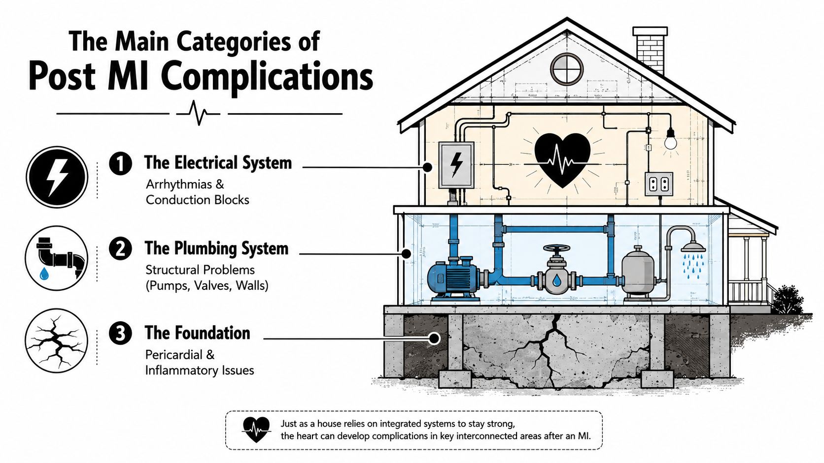
Think of the heart as a house damaged by a major storm. The storm has passed, but the wiring, pipes, walls, pump, and foundation may need inspection. This analogy doesn't replace medical language, but it gives you a useful mental map.
Electrical wiring problems
The heart's electrical system controls rate and timing. Arrhythmias include rhythms that are too fast, too slow, or irregular, while conduction blocks interrupt the signal as it travels through the heart.
You might feel this as racing, pounding, fluttering, pauses, dizziness, or fainting. Some rhythm changes are temporary, but dangerous ventricular rhythms and significant conduction disturbances can reduce blood flow quickly. Arrhythmias and conduction disturbances can occur in up to 90% of patients after MI, according to a review in the MSD Manual.
Plumbing problems
The coronary arteries are the heart's fuel pipes. A new blockage, a clot, or a problem with the treated artery can reduce blood flow again, causing recurrent ischemia or another infarction.
The everyday example is new pressure in the chest during rest, especially when it feels similar to the original heart attack. That symptom should never be explained away as indigestion without medical assessment.
Wall and valve damage
A heart attack can weaken heart muscle. In uncommon cases, the damaged tissue can tear, causing a ventricular septal rupture, papillary muscle rupture with sudden severe mitral regurgitation, or free-wall rupture.
These are structural emergencies. A new murmur, sudden breathlessness, collapse, or rapidly worsening weakness may reflect a change in how blood moves through the heart. Mechanical complications occur in less than 1% to 6.2% of acute MI patients and account for 14% to 26% of infarct-related mortality, according to this European Society of Cardiology teaching source.
A failing pump
When injured heart muscle can't circulate blood effectively, heart failure may develop. Fluid can collect in the lungs or legs, making ordinary activity harder.
A person may notice increasing breathlessness, difficulty lying flat, swelling, rapid weight change, or unusual exhaustion. Heart failure can appear soon after the MI or become a longer-term issue as the heart remodels.
An inflamed lining
The pericardium is the protective lining around the heart. Inflammation after an MI, called post-MI pericarditis, may cause sharp chest pain that changes with breathing or position.
Because chest pain after an MI has many possible causes, don't self-diagnose it. A plain-language explanation of this condition is available in this guide to pericarditis.
Clot-related problems
A weakened or poorly moving section of the left ventricle can allow a clot, called an LV thrombus, to form. If part of that clot travels, it can cause an embolism or stroke.
Sudden one-sided weakness, facial drooping, confusion, or trouble speaking is an emergency. Blood-thinning treatment may be appropriate for some patients, but never start, stop, or adjust it without your clinician's direction.
When Each Complication Typically Shows Up
Timing helps, but it doesn't provide a guarantee. A complication can appear outside its usual window, so new or worsening symptoms still matter even when you're months or years beyond the original event.
The first 24 hours
The earliest period is when electrical instability and impaired pumping receive the most attention. Ventricular tachyarrhythmias can cause sudden cardiac death, while severe pump failure can lead to cardiogenic shock. Continuous monitoring helps the hospital team identify changes before a patient can describe them.
For STEMI, guidance recommends ECG monitoring for arrhythmias and ST-segment changes for at least 24 hours after symptom onset. Longer observation should be considered with higher-risk findings such as hemodynamic instability, major arrhythmias, LVEF below 40%, failed reperfusion, important additional stenoses, or PCI complications, according to the European Heart Journal guidance.
Days two through seven
Structural problems may become apparent after the initial treatment, especially when the infarct damaged the full thickness of the heart muscle. Ventricular septal rupture, papillary muscle rupture, and acute mitral regurgitation can produce sudden breathlessness, low blood pressure, a new murmur, or collapse.
Post-MI pericarditis and early heart failure may also enter the picture. Mechanical complications remain uncommon in the reperfusion era. Contemporary estimates place ventricular septal rupture at 0.17% to 0.91%, papillary muscle rupture at 0.05% to 0.26%, and free-wall rupture at 0.01% to 0.52%, as summarized in the Annals of Cardiothoracic Surgery review.
Weeks two through six
This is often when people feel caught between hospital care and ordinary life. The heart may continue to remodel, and clinicians may reassess pumping function, symptoms, medications, and rhythm. LV thrombus risk depends on the size and location of the infarct, how well the ventricle contracts, and other clinical findings.
A new symptom at three weeks still deserves attention. Don't use the calendar to dismiss chest discomfort, swelling, breathlessness, fainting, or a sustained irregular heartbeat. Even questions about follow-up blood tests can be useful, including what clinicians expect from troponin after a heart attack.
Beyond six months
Long-term risk doesn't vanish when the scar has matured. In a population study of 433,361 heart-attack patients, heart failure occurred in 29.6%, renal failure in 27.2%, atrial fibrillation in 22.3%, severe bleeding in 19.0%, and a subsequent MI in 7.1% over follow-up. All-cause mortality was the most frequent outcome at 37.8%, according to the ESC 2023 post-MI outcomes abstract.
Those figures aren't a prediction of your personal future. They explain why follow-up, medication review, rehabilitation, and attention to new symptoms remain important long after discharge.
Common Sensations Versus True Warning Signs
Recovery can include sensations that are uncomfortable but not automatically dangerous. Mild fatigue, incision soreness after a procedure, brief lightheadedness after standing too quickly, and an occasional flutter may occur during the adjustment period.
The key question is pattern and severity. A brief sensation that resolves and doesn't recur is different from a symptom that lasts, intensifies, appears at rest, or arrives with fainting or breathlessness.
Symptoms that should prompt emergency action
Call emergency services for:
- Chest pressure at rest: Especially if it resembles the original MI, spreads to the arm, jaw, back, or shoulder, or comes with sweating or nausea.
- Sudden severe breathlessness: This is more concerning when it occurs at rest, wakes you from sleep, or prevents you from speaking comfortably.
- Fainting or near-fainting: A collapse can reflect a dangerous rhythm, low blood pressure, or reduced cardiac output.
- Rapid sustained palpitations: A fast rhythm that continues, particularly with chest pain, dizziness, weakness, or breathlessness, needs urgent assessment.
- Sudden swelling or marked fluid symptoms: New, rapidly worsening swelling or breathlessness may indicate heart failure.
- Coughing up blood: This requires prompt emergency evaluation.
- Stroke symptoms: One-sided weakness, facial drooping, confusion, or slurred speech should be treated as an emergency.
If anxiety is amplifying chest sensations, that still doesn't make the symptom imaginary. A resource on anxiety and chest pain can help explain how stress affects the body, but after an MI, new chest pressure should be assessed medically rather than attributed to anxiety.
A late-night decision
Suppose you wake at 11 p.m. with a flutter. First, sit down safely and notice whether it is continuing. If it's brief and you feel completely well, write down the time, duration, pulse if available, and any associated symptoms, then contact your clinician for advice if it repeats.
If the flutter continues or comes with chest pressure, faintness, severe breathlessness, or weakness, skip the app and call emergency services. You know what feels different in your body. No algorithm can replace that judgment or an in-person assessment. When you're unsure, calling is the safer choice. The cardiac event symptoms guide can also help you prepare questions for your care team.
Get your ECG checked by certified experts within minutes on the Qaly app.


How Doctors Check for Complications and What Those Tests Mean
Each test answers a different question. An ECG is a snapshot of electrical activity, while telemetry watches the rhythm continuously in a monitored setting. The team may look for recurrent ischemia, abnormal rhythms, conduction blocks, or changes that suggest the heart is under strain.
An echocardiogram uses sound waves to show how the chambers and valves move. It can estimate pumping function, identify fluid around the heart, detect a septal defect, assess mitral regurgitation, and show whether part of the ventricle is moving poorly. If the result is unclear, cardiac MRI can provide a more detailed view of scar, inflammation, tissue injury, and ventricular structure.
What blood tests add
Troponin reflects heart muscle injury, but the result has to be interpreted alongside symptoms, ECG findings, kidney function, and the clinical timeline. BNP or a related natriuretic peptide can support the assessment of heart failure, especially when breathlessness has more than one possible cause.
These tests don't function like pass-or-fail exams. An abnormal value tells the clinician what deserves closer attention, not automatically what the final diagnosis is.
Why follow-up testing matters
Doctors may repeat an echocardiogram because the heart's pumping function can change during healing and remodeling. They may order rhythm monitoring because intermittent AF, ventricular arrhythmias, or AV block can appear after discharge, even when a short office ECG looks normal.
Stress testing is considered in selected situations to assess how the heart responds to exertion and whether symptoms suggest inducible ischemia. It isn't appropriate for every patient at every stage, so ask what clinical question the test is meant to answer.
Ask directly: “What complication are you checking for, and what result would change my treatment?”
That question makes the process more transparent. It also helps you understand whether a test is looking at the artery, the pump, the valves, the rhythm, inflammation, or medication safety.
At home, wearable ECG recordings can add context when symptoms are intermittent, but they don't assess blocked arteries, pumping strength, or structural tears. They're one piece of information to bring to the clinical team, not a replacement for a 12-lead ECG, imaging, or emergency evaluation.
Tracking Your Heart Rhythm at Home with a Wearable ECG
A short office ECG can look normal because the rhythm problem wasn't happening during the appointment. Wearable ECG devices are useful for capturing an intermittent event, especially when you record it while symptoms are present.
A single-lead tracing can show rate and rhythm patterns. Depending on the device and interpretation, it may help identify irregular rhythms, pauses, or a rhythm that warrants professional review. It generally can't show the full information available from a 12-lead ECG, diagnose a blocked artery, measure heart structure, or prove that chest pain is harmless.

Build a useful record
Symptom-triggered recordings are valuable, but they can miss silent AF, nonsustained ventricular tachycardia, or AV block. That's why a wearable should support, not replace, the monitoring plan your cardiologist recommends.
Keep a simple log with:
- The moment it started: Include the date and approximate time.
- What you felt: Note fluttering, racing, pauses, dizziness, chest discomfort, or breathlessness.
- How long it lasted: Record whether it stopped on its own or continued.
- What you were doing: Resting, walking, climbing stairs, lying down, or feeling stressed can provide useful context.
- Medication details: Record missed doses, recent changes, and symptoms that began after a new medicine.
- Available ECG details: Heart rate, rhythm regularity, and PR, QRS, or QTc intervals may help clinicians review trends when the device provides them.
A frightening label on a screen isn't a diagnosis. Save the recording, write down how you felt, and send it through the communication method your clinician has approved. If the reading appears abnormal and you also have emergency symptoms, don't wait for a report or message response.
Some patients also explore lifestyle resources discussing evidence-based HRV optimization. Heart-rate variability can be influenced by sleep, stress, illness, medications, and recovery, but an HRV score shouldn't overrule symptoms or your cardiologist's advice.
A human-reviewed ECG service may provide interval measurements and narrative context that are easier to share at an appointment. Still, no wearable interpretation can rule out a heart attack, evaluate sudden collapse, or safely manage severe breathlessness. Emergency symptoms require emergency care first.
Living Well After MI Without Losing Your Mind
You can't control every post-MI complication, but you can create conditions that make problems easier to prevent, detect, and treat. Take prescribed medicines consistently, and report side effects rather than stopping a medication on your own. Sudden changes to antiplatelet, anticoagulant, blood pressure, or rhythm medicines can create avoidable risk.
Cardiac rehabilitation gives you a supervised way to rebuild strength and confidence. Ask when you can begin, which activities are appropriate, and what symptoms should make you stop. Gradual activity supports recovery without turning every walk into a test of willpower.
Your daily choices connect to the systems described earlier:
- Electrical stability: Sleep, medication consistency, and reporting palpitations help your team understand rhythm changes.
- Pump protection: Follow-up imaging, gradual activity, and attention to breathlessness or swelling support early recognition of heart failure.
- Plumbing protection: Take artery-protective medicines as directed and discuss blood pressure and cholesterol goals with your clinician.
- Clot prevention: Ask exactly how to take any prescribed blood thinner and which bleeding symptoms require a call.
- Emotional recovery: Stress management, supportive relationships, and counseling can reduce the sense that every heartbeat is a threat.
Alcohol, sleep, work, driving, and sexual activity all deserve specific guidance rather than guesswork. If an instruction seems inconsistent, a side effect feels unacceptable, or your concerns aren't being addressed, ask for clarification or seek a second opinion. Skepticism can be constructive when it leads to better questions and shared decisions.
Recovery rarely follows a straight line. A difficult day is information, not failure. Build a small, trusted team that includes your cardiologist, primary clinician, family, and a reliable ECG-reading service when appropriate. Keep your records, bring questions, and speak up when something changes.
If you're using a wearable ECG after an MI, start a symptom-and-reading log today and share it at your next appointment. For an added layer of rhythm context, try Qaly's human-reviewed ECG analysis, then use the report to have a more focused conversation with your cardiologist.
Unsure about an ECG? Certified experts review it within minutes, day or night.


