Key Takeaways
Hello Heart Hero. A heart echocardiogram is basically an ultrasound for your heart. It creates live, moving pictures of it without any invasive procedures. This safe, painless test uses sound waves to show your doctor exactly how well your heart is pumping blood and whether its structure looks healthy.
Your Guide to Understanding Your Heart Health
If your doctor has brought up a heart echocardiogram, it’s natural to feel a bit of curiosity, and maybe some concern. We get it. Medical tests can feel like a maze, especially when you’re just trying to get clear answers in a healthcare system that can sometimes feel impersonal.
This guide is meant to be a friendly companion on that journey. We'll walk you through what an echocardiogram is, using simple analogies instead of dense medical jargon. Our goal is to swap any uncertainty you have with clarity and confidence.
Why This Test Is a Powerful Tool
Think of this test not as something to worry about, but as a window into your heart’s health. It gives a clear, real-time picture that helps you and your doctor make the best decisions for your future. It's a fantastic, proactive step.
Here’s a quick rundown of why it's so helpful:
- It is non-invasive and painless: No needles or cuts are involved in a standard echo.
- It provides live images: Unlike a static photo, an echo shows your heart in action, revealing how it moves and pumps.
- It is a radiation-free procedure: The test uses sound waves, making it incredibly safe, even if you need it more than once.
Understanding the "why" behind the test is just as important as the "what". Seeing the echo as one piece of your overall health journey helps put things in perspective.
Think of an echocardiogram as giving your doctor a direct look "under the hood" of your heart. It allows them to check the engine's performance, look for worn-out parts, and ensure everything is running smoothly, all without taking anything apart.
While this test gives an amazing, in-depth look at your heart's mechanics, you might be curious about what else you can do. It's empowering to learn how to check heart health at home to complement the care you get from your doctor. Let’s explore this together, so you feel fully prepared for your appointment.
What a Heart Echocardiogram Really Is
Let's clear the air and demystify the heart echocardiogram. We know medical terms can sound intimidating, but this one is much simpler than it seems. At its core, an echocardiogram is just a heart ultrasound. It’s a way for your doctor to get a live, moving picture of your heart without any invasive procedures.
Think of it like a submarine using sonar to map the ocean floor. The sub sends out sound waves that bounce off objects and return, creating a detailed map of what’s below. An echocardiogram works on the exact same principle, but for your heart. It uses safe, high-frequency sound waves that you can’t hear or feel.
This completely painless process allows your doctor to see your heart in action, giving them a clear view of its function without any guesswork.

How It Creates a Picture of Your Heart
During the test, a small handheld device called a transducer is gently placed on your chest. This little wand sends out those sound waves, which travel through your skin and tissues. When they reach your heart, they bounce off its various parts like the chambers, the valves, and the muscular walls.
Those returning sound waves are like echoes. The transducer picks them up, and a computer immediately translates them into detailed, real-time images on a screen. It’s like watching a live video feed of your own heart beating.
This visual information is incredibly valuable. It lets your doctor check several key aspects of your heart's health:
- Pumping Action: They can see how well your heart chambers are contracting and squeezing blood out to your body.
- Size and Shape: The test shows the size of your heart and the thickness of its walls, flagging any enlargement or other issues.
- Valve Function: Doctors can watch your heart valves open and close with each beat to make sure they aren't leaky or too narrow.
In essence, an echocardiogram provides a direct, non-invasive look at your heart's mechanical performance. It’s not about interpreting electrical signals or looking at a static image; it’s about observing the living, beating organ as it does its job.
This technology is a crucial component of cardiac care due to its safety and effectiveness. It has become a widely trusted and utilized tool in the field.
Differentiating From Other Heart Tests
It's easy to get heart tests mixed up, especially since many have similar-sounding names. Just remember, an echocardiogram focuses on the heart’s structure and blood flow, the "plumbing and mechanics" of the organ.
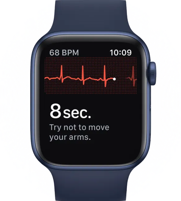
This is very different from a test that measures the heart's electrical activity. To get a full picture, understanding what an ECG test involves can be helpful, as an ECG tracks the electrical impulses that trigger your heartbeat. Think of it as checking the "wiring."
Ultimately, a heart echocardiogram gives you and your doctor a clear, reliable picture to work from. It replaces uncertainty with direct visual evidence, empowering you with knowledge about your own body and helping guide the best path forward for your health.
Why Your Doctor Recommended This Test
Hearing your doctor wants you to have a heart test can be unsettling. It’s completely normal to have questions or feel a little anxious. Let's start by saying that an echocardiogram is an incredibly common, safe, and proactive way for your doctor to get a better look at your heart.
Your doctor likely recommended this test for a specific reason, either to figure out what’s causing a symptom you’ve been feeling or to keep an eye on a known condition. Think of it as a detailed health check for your heart. It moves beyond guesswork to give your medical team concrete information to create the best care plan for you.
Investigating Common Symptoms
Often, an echocardiogram is the next logical step when you report certain symptoms. It’s a powerful tool for connecting the dots between how you're feeling and what's actually happening inside your chest, helping to find the root cause of issues that can sometimes feel vague or concerning.
Some of the most common reasons a doctor will order an echo include:
- Shortness of Breath: If you find yourself getting breathless from simple activities, an echo can check if your heart is pumping blood as efficiently as it should. You can learn more about what causes shortness of breath in our detailed guide.
- Chest Pain: While not all chest pain is heart-related, an echocardiogram is a great way to rule out or identify structural problems that could be the culprit.
- Dizziness or Fainting: These symptoms can sometimes be caused by irregular blood flow or issues with your heart valves, both of which are clearly visible on an echo.
- Irregular Heartbeat: If you've felt palpitations or a fluttering sensation, this test can show if a structural issue with your heart is the cause.
- Swelling in the Legs: Fluid retention, also known as edema, can be a sign that your heart isn’t pumping as strongly as it needs to, a condition an echo can easily assess.
Diagnosing and Monitoring Heart Conditions
Beyond just looking into symptoms, an echocardiogram is essential for diagnosing and managing a wide range of specific heart conditions. It gives your doctor the detailed visual evidence needed to confirm a diagnosis and understand its severity.
This non-invasive test is a fundamental aspect of modern cardiology due to its importance and effectiveness. The standard transthoracic echocardiogram (TTE) is especially notable for being a safe and reliable first-line tool in assessing heart health.
This test is particularly good for several key things:
- Assessing Heart Valve Problems: It can spot if your heart valves are too narrow (stenosis) or if they are leaking (regurgitation).
- Detecting Heart Failure: It measures how well your heart muscle is pumping blood, which is a critical piece of information for diagnosing heart failure.
- Evaluating Damage from a Heart Attack: After a heart attack, an echo can reveal which areas of the heart muscle have been damaged.
- Identifying Congenital Heart Defects: The test is used to find structural heart problems that have been present since birth.
- Checking for Blood Clots or Tumors: It can show if there are any abnormal growths inside your heart's chambers.
An echocardiogram is more than just a diagnostic snapshot. It's an ongoing tool for partnership between you and your doctor, allowing for informed, collaborative decisions about your health journey.
It can also be used to track how well treatments like medication or surgery are working, or to check on your heart's health before a major non-cardiac surgery. In every case, the goal is the same: to give you and your doctor the clearest picture possible so you can feel empowered and confident in your care.
Exploring the Different Types of Echocardiograms
It helps to know that "heart echocardiogram" isn't a one-size-fits-all term. Think of it more like a toolkit, where your doctor picks the right tool for the specific job at hand. Depending on what they need to see, there are several different types of echocardiograms, each giving a unique view of your heart.
Understanding these different types can help you feel more prepared and less in the dark about your own health. You'll have a better idea of what to expect and why your doctor chose a particular test for you. Let's walk through the most common ones.
The Standard Transthoracic Echocardiogram (TTE)
This is the test most people picture when they hear "echocardiogram." The Transthoracic Echocardiogram, or TTE, is the standard, completely non-invasive procedure we've been talking about. "Transthoracic" is just a medical way of saying "across the chest."
During a TTE, the sonographer simply moves the transducer over different parts of your chest and upper abdomen to get various pictures of your heart. It’s painless, totally safe, and gives a fantastic overall look at your heart's structure and function. This is almost always the first type of echo a person will have.
The Closer Look with a Transesophageal Echocardiogram (TEE)
Sometimes, your doctor needs a clearer, more detailed picture, especially of the back of the heart. Your ribs and lungs can get in the way of the sound waves during a standard TTE, creating some fuzzy spots. For a high-definition view, they might turn to a Transesophageal Echocardiogram (TEE).
With a TEE, a much smaller transducer is put on the end of a thin, flexible tube. This tube is gently guided down your throat into your esophagus (the tube leading to your stomach). Since your esophagus sits right behind your heart, this placement gives incredibly clear, unobstructed images.
Don't worry, you’ll be given medication to help you relax and to numb your throat, so you won't feel any pain. A TEE is often the go-to for:
- Getting a better look at the aorta or heart valves.
- Checking for blood clots inside the heart's upper chambers.
- Guiding certain heart procedures in real-time.
Seeing Your Heart Under Pressure with a Stress Echocardiogram
A standard echo shows how your heart works while you’re resting. But what about when it’s working hard? A Stress Echocardiogram is designed to answer that exact question. It's a two-part test that compares images of your heart at rest with images taken right after it's been stressed.
First, you'll have a resting TTE. Then, you’ll get your heart rate up, usually by walking on a treadmill or riding a stationary bike. If you can't exercise, you might be given a medication that makes your heart beat faster, mimicking the effects of physical activity.
Right after the stress part, you'll lie down again so the sonographer can take a second set of images. By comparing the "before" and "after" pictures, your doctor can see how your heart muscle and blood flow respond to the demand. This is an excellent way to look for signs of coronary artery disease.
An echocardiogram serves several key purposes, from diagnosing initial symptoms to monitoring long-term heart conditions. It empowers both you and your doctor with clear visual information.
This infographic neatly summarizes the core purposes of a heart echocardiogram.

As you can see, the test is a versatile tool used to diagnose, monitor, and evaluate treatments, making it central to comprehensive heart care.
Advanced and Specialized Echocardiograms
Technology is always moving forward, and echocardiography is no exception. Beyond these main types, there are more advanced variations that give us even greater detail. A 3D echocardiogram, for instance, creates a stunningly detailed three-dimensional model of your heart, which can be invaluable for planning surgery.
Another specialized type is intracardiac echocardiography (ICE). This advanced technique involves imaging the heart from the inside during complex procedures. It’s becoming essential for guiding treatments for conditions like atrial fibrillation and valve repair.
Given that cardiovascular disease led to 931,578 deaths in the U.S. in 2024, an increase of 4% from the year before, having such precise tools is incredibly important. You can find out more about advancements in intracardiac echocardiography from this market report.
Each type of echocardiogram offers a different piece of the puzzle. By choosing the right one, your doctor gets the exact information needed to support your heart health journey with confidence and clarity.
Preparing for Your Echocardiogram Appointment
It's completely normal to feel a little nervous before any medical test. The good news? Prepping for a standard transthoracic echocardiogram (TTE) is incredibly simple. There are no complicated steps, which helps take the edge off any pre-test jitters.
You can eat and drink normally before your appointment. No fasting is required, so feel free to have your usual breakfast or lunch. This simple detail often makes the whole experience feel less like a major medical event and more like a routine check-in.
What to Wear and Expect on the Day
To keep things running smoothly, it's best to wear comfortable, loose-fitting, two-piece clothing. Think a shirt with pants or a skirt. This is just a practical matter, as you'll need to undress from the waist up. You'll be given a gown to wear for privacy and comfort.
The test itself is very straightforward and usually takes between 30 and 60 minutes. It's a quiet, calm procedure where you just get to lie back and relax.
Here’s a step-by-step look at what will happen once you’re in the exam room:
- Getting Comfortable: You'll be asked to lie on an examination table, typically on your left side. This position helps bring your heart a bit closer to the chest wall, which allows for clearer images.
- Placing Electrodes: A trained technician, called a sonographer, will attach small, sticky patches called electrodes to your chest. These are connected to a machine that tracks your heart's electrical rhythm during the test, but you won't feel a thing from them.
- Applying the Gel: Next, the sonographer will squeeze a clear, water-based gel onto your chest. It might feel a bit cool at first, but it's harmless and wipes off easily afterward. This gel is key because it helps the sound waves travel from the probe into your body.
During the Echocardiogram Procedure
Once the gel is on, the sonographer will gently press a small, handheld device called a transducer firmly against your skin. They'll move it around to different spots on your chest to capture various views and angles of your heart. Think of it like a photographer moving around to get the perfect shot.
You might be asked to hold your breath for a few seconds here and there or to shift your position slightly. This just helps the sonographer get the sharpest pictures possible. While you may feel some light pressure from the transducer, the procedure should not be painful. If you feel any discomfort at all, don't hesitate to let the sonographer know.
Your only job during a heart echocardiogram is to relax. The sonographer handles all the technical stuff, letting you simply lie back while they gather the important information your doctor needs.
After the test, the sonographer will wipe the gel off, and you can get dressed and go about your day. There’s no recovery time needed. This is a great moment to gather your thoughts before your follow-up with the doctor. It's always helpful to have some questions ready to make that conversation as productive as possible. For ideas, you might want to review these questions to ask your cardiologist so you feel fully prepared to discuss the results.
How to Interpret Your Echocardiogram Results
Once your heart echocardiogram is finished, you don't just get a handful of pictures. Instead, a cardiologist, a doctor specializing in the heart, will carefully analyze the moving images and put together a detailed report. This report then goes to your primary doctor, who will sit down with you to go over what it all means.
Think of this document as a comprehensive inspection report for your heart. Its job is to give your doctor clear, actionable information to guide your care, so you don't have to figure it out on your own.

What the Report Actually Measures
An echocardiogram report dives into several key factors, and each one offers a different clue about your heart’s overall condition. Your doctor will translate these measurements into a meaningful story about how your heart is functioning day-to-day.
Here are some of the main things the report will cover:
- Heart Chamber Size: The report will note the dimensions of your heart's four chambers. An enlarged chamber can be a sign that the heart is working too hard to pump blood.
- Heart Wall Thickness: It also measures the thickness of the muscular walls. Walls that are too thick or too thin can point toward different types of heart disease.
- Pumping Strength: This is one of the most important takeaways from the test, showing how effectively your heart pushes blood out with each beat.
Understanding Ejection Fraction (EF)
A critical number you'll see in your report is the ejection fraction, or EF for short. This measurement tells you what percentage of blood is pumped out of your heart's main pumping chamber (the left ventricle) with each squeeze. It’s a direct indicator of your heart’s pumping power.
A normal EF is typically between 50% and 70%. This means a healthy heart pushes out a little over half the blood in the ventricle with every beat. It doesn't pump out 100% because it needs some blood left inside to maintain its shape.
Your ejection fraction is a key metric for heart health. A lower-than-normal EF can be one of the first signs of conditions like heart failure, which is when the heart muscle isn't pumping as well as it should.
A low EF might sound alarming, but it gives your doctor vital information to create an effective treatment plan. If you're looking to learn more about this condition, our guide on understanding heart failure and what it looks like on your ECG provides some helpful context.
Checking Your Heart Valves and Structure
Beyond the pumping action, the report gives a detailed look at your heart's valves. The cardiologist checks if they are opening and closing properly, noting any signs of:
- Stenosis: This is when a valve gets stiff or narrowed, making it harder for blood to flow through.
- Regurgitation: This happens when a valve doesn’t seal tightly, letting blood leak backward.
Finally, the report will highlight any other structural issues. This could be anything from damage from a past heart attack to fluid buildup around the heart or even a defect you were born with. Your doctor will connect all these dots to give you a complete picture of your heart health, explaining exactly what the results mean for you and what the next steps are.
Your Echocardiogram Questions, Answered
Heading into any heart procedure, it's completely normal to have a few questions. Feeling informed and prepared is one of the best ways to ease any anxiety you might be feeling. Let's walk through some of the most common questions people have about echocardiograms.
Is an Echocardiogram Painful?
Not at all. A standard transthoracic echocardiogram is completely painless. You might notice the gel on your chest feels a bit cool, and you'll feel some light pressure as the sonographer guides the transducer over your skin, but there shouldn't be any pain.
Is There Any Radiation Exposure?
This is an excellent and important question. An echocardiogram does not use any radiation. It works with safe, high-frequency sound waves, the same kind of ultrasound technology used to see babies during pregnancy. This makes it a very safe test, even if you need to have it done more than once.
How Long Does the Test Take?
You can expect a standard echocardiogram to take between 30 to 60 minutes. The exact duration really just depends on how easily the sonographer can get clear images of your heart. It’s a pretty quick and efficient process.
The whole experience is designed to be as simple and comfortable for you as possible. Your only job is to lie back and relax while the technology does its thing, gathering all the necessary information for your doctor.
When Will I Get My Results?
While the images are captured live during the test, they need to be carefully analyzed by a cardiologist. This specialist reviews the images and creates a detailed report for your doctor, which usually takes a few days. Your doctor will then schedule a time to go over the findings with you.
Get expert analysis of your wearable ECGs in minutes and the peace of mind that comes with it.


.png)
.png)