

Key Takeaways
- A normal ECG reflects a healthy and well-timed heart rhythm. It includes consistent P waves, narrow QRS complexes, and regular intervals like the PR and QT intervals. These patterns show that the heart is beating in a steady and efficient way.
- What is considered normal can differ from person to person. For example, athletes often have lower heart rates, and women may have slightly longer QT intervals than men.
- Common ECG variations can still be healthy. Sinus arrhythmia, early repolarization, and occasional extra beats such as PACs or PVCs are frequently seen in healthy individuals and are not always a cause for concern.
User Profile
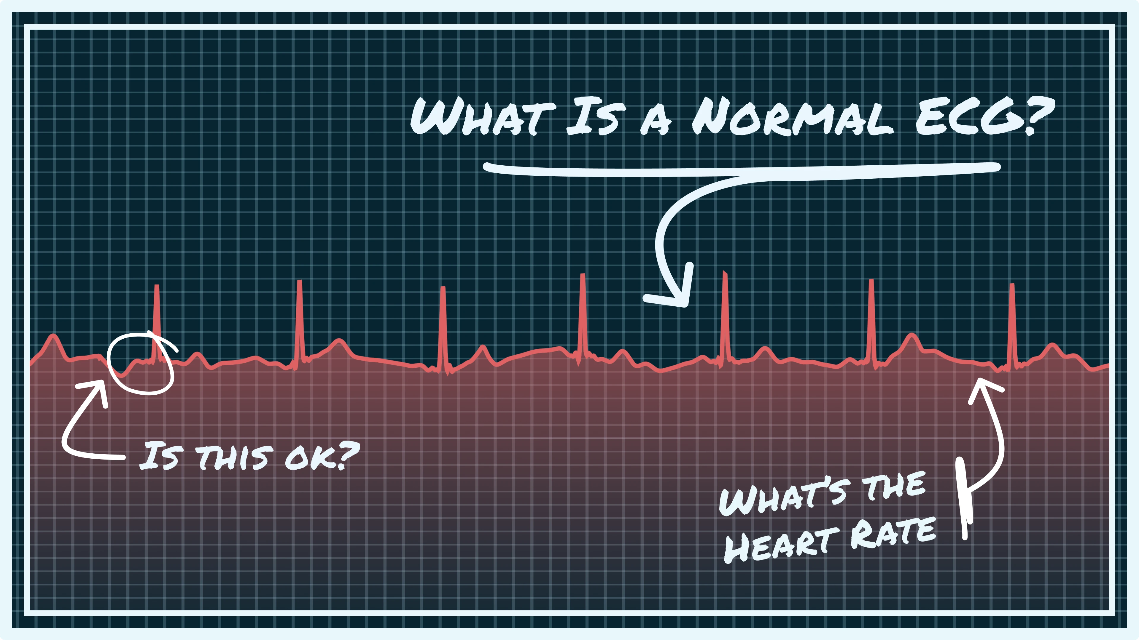
Understanding a normal electrocardiogram (ECG) reading is like figuring out your heart's electrical signals. An ECG turns the heart's electrical activity into a visual chart, helping doctors check its rhythm and function. Let's look at the main parts of a normal ECG and what they mean.
The Basic Building Blocks: Waves, Complexes, and Intervals
A normal ECG has distinct visual patterns made up of waves, complexes, and intervals. The P wave shows the electrical activation of the atria, marking the start of a heartbeat. This is followed by the QRS complex, a more prominent spike that signals the ventricles contracting. The T wave represents the ventricles' repolarization, their recovery period before the next beat. The time between these components, like the PR interval and QT interval, provides crucial details about the timing of the heart's electrical conduction.
What Does a Normal ECG Reading Look Like?
A normal ECG shows these elements in a consistent, rhythmic pattern. The heart rate should be within a healthy range (typically 60-100 beats per minute in adults). A P wave should always come before each QRS complex, showing a smooth electrical flow from the atria to the ventricles. The QRS complex should be narrow, indicating effective ventricular contraction. Consistent intervals between components further demonstrate a healthy electrical system.
Normal ECG Values and Their Significance
A normal ECG reading is key to understanding heart health. Typically, the P wave duration should be less than 130 ms in adults, while the QRS duration should be less than 110 ms for men and less than 100 ms for women. These values indicate the speed of electrical conduction in different parts of the heart. Research using ECG data from over 152,000 people showed a tachycardia (heart rate > 100 bpm) prevalence of 0.71% and bradycardia (heart rate < 50 bpm) prevalence of 3.39% in adults aged 18 to 65. This data helps define normal heart rate ranges, recognizing that variations can occur depending on factors like age and health conditions. It's important to remember that minor variations within the normal range are possible and don't necessarily signal a problem. A comprehensive evaluation considers individual factors and the overall clinical picture.
The Numbers Behind Normal: Essential ECG Measurements
An electrocardiogram (ECG) provides a visual representation of your heart's electrical activity. Beyond the recognizable peaks and valleys, crucial numerical measurements provide deeper insights into heart function. These values determine whether your heart's electrical activity is within the normal range.
Heart Rate: Understanding Your Heart's Rhythm
The heart rate, a fundamental ECG measurement, indicates the number of times your heart beats per minute (bpm). A normal resting heart rate for adults typically falls between 60 and 100 bpm. This number signifies the complete electrical cycles your heart performs to pump blood throughout your body.
Several factors can influence heart rate, including age, physical fitness, and emotional state. Athletes, for instance, often have lower resting heart rates due to their conditioned cardiovascular systems.
Intervals and Durations: Precision Timing
The timing of the heart's electrical signals is just as important as the rate. These timings, measured as intervals and durations on the ECG, provide critical information about heart function.

The PR interval, for example, measures the time it takes for the electrical impulse to travel from the atria to the ventricles. A normal PR interval typically ranges from 120 to 200 milliseconds (ms). The QRS duration, representing ventricular depolarization, is usually less than 100 ms. These precise timings reveal the efficiency of the heart's conduction system.
Amplitude: Strength of the Electrical Signals
The amplitude, or height, of the ECG waves is another important factor. While less strictly defined than heart rate or intervals, amplitude offers insights into the strength of the electrical signals. Abnormally high or low amplitudes can suggest underlying heart conditions, emphasizing the importance of considering amplitude in conjunction with other measurements.

Interpreting ECGs relies heavily on understanding normal values, considering age and gender variations. Further research has shown that guidelines have evolved to accommodate these factors, improving our understanding of how age and gender influence cardiac electrophysiology. For adult males, a QRS duration up to 110 ms has been considered normal.
Normal ECG Values
The table below shows the typical ranges for essential ECG measurements in healthy adults.
This table summarizes the accepted normal ranges for key ECG parameters. Significant deviations from these ranges may warrant further investigation by a healthcare professional. A prolonged QT interval, for example, can increase the risk of certain arrhythmias. Comparing an individual's ECG measurements against these established norms helps healthcare providers identify potential abnormalities and recommend appropriate follow-up care.
Why Your Normal Isn't My Normal: Age and Gender Factors
A "normal" ECG reading isn't one-size-fits-all. Just like other health metrics, what's considered normal on an ECG can vary based on age and gender. What's normal for a 20-year-old woman will likely look different for a 70-year-old man. Understanding these differences is key for accurate interpretation.
The Impact of Age on ECG Readings
Our hearts change as we age, and these changes show up on our ECGs. For instance, heart rate tends to slow down, and the electrical signals might not travel as efficiently. This can result in slightly longer PR intervals and QRS durations in older adults. The T wave, another part of the ECG, can also change shape with age.
Gender Differences in ECGs
Gender also influences a normal ECG. Women often have shorter QRS durations and longer QT intervals than men. This difference is likely due to hormones and variations in heart size and structure. Knowing these gender-specific norms helps prevent misinterpretations. A slightly longer QT interval might be perfectly normal for a woman but could raise a flag for a man.
Putting It All Together: Context Is Key
These variations underscore the importance of context in ECG interpretation. Experts who review thousands of ECGs every year know that strict numerical limits aren't always enough. They also consider the patient's age, gender, medical history, and overall health.
This personalized approach is especially valuable for services like Qaly, which provides human-reviewed ECG analysis. Qaly considers these individual factors to ensure accurate and personalized insights. A slightly prolonged QRS complex could be normal in an older person, while the same result might be flagged as potentially abnormal in a younger person. This contextualized interpretation can help prevent unnecessary worry and ensure appropriate care. It empowers users to track their heart health and discuss results with their doctors. This collaborative approach provides a clearer understanding of ECG results and guides appropriate care.
When Abnormal Isn't Actually Abnormal: Common ECG Variations
Not every unusual blip on an ECG signals a serious problem. Many variations, while appearing out of the ordinary, are actually benign adaptations reflecting normal physiological processes. This means what might initially seem concerning can be perfectly healthy. Understanding these variations is crucial for avoiding unnecessary anxiety and redundant testing.
Breathing and Your Heart Rhythm: Respiratory Sinus Arrhythmia
One common variation is respiratory sinus arrhythmia, where the heart rate subtly changes with breathing. This is particularly noticeable in younger individuals and is considered a sign of healthy heart-lung interaction.
.webp)
During inhalation, the heart rate slightly increases, and during exhalation, it slows down. This fluctuation is driven by changes in vagal nerve activity and is entirely normal.
The Athlete's Heart: Training-Induced Changes
Athletic training can also reshape ECG readings. Athletes often develop bradycardia, a slower resting heart rate, sometimes dipping below 60 beats per minute.
This is because their hearts become more efficient, needing fewer beats to circulate blood. Additionally, other ECG changes, like slight increases in the size of certain heart chambers, can be seen in highly trained athletes. These adaptations reflect peak cardiovascular conditioning, not pathology.
Benign ECG Patterns: Early Repolarization and Juvenile T Waves
Certain ECG patterns, although they may differ from the standard definition of "normal," are often observed and are harmless. Early repolarization, which involves specific alterations in the ST segmentand T wave, is one such example. Unfortunately, devices like the Apple Watch cannot accurately track changes in the T wave and ST segments.
Similarly, juvenile T-wave patterns, frequently seen in children and adolescents, exhibit distinct T-wave shapes that may appear unusual but are a normal aspect of development.
Distinguishing Normal from Abnormal: The Cardiologist's Expertise
Experienced cardiologists understand the nuances of ECG interpretation. They consider not just the readings but also the patient's age, medical history, lifestyle, and symptoms. ECG is a widely used diagnostic tool, with over 300 million procedures performed annually worldwide.
Interestingly, a study found that about 77.7% of individuals without prior heart issues had a normal ECG, while 16.8% had at least one minor abnormality. Find more detailed statistics here. This underscores the importance of expert interpretation in distinguishing harmless variations from true abnormalities.
For example, occasional premature atrial contractions (PACs) or premature ventricular contractions (PVCs) – extra heartbeats – are often benign, especially without other symptoms. Services like Qaly leverage expert analysis to provide personalized insights into ECG readings, helping users understand their heart health without undue concern. This approach helps reassure patients worried about minor ECG variations and ensures that potential issues are addressed appropriately.

Red Flags: When ECG Patterns Signal Potential Problems
After exploring normal ECG readings and common variations, it's important to understand which patterns might indicate a problem. These red flags require careful evaluation by healthcare professionals, and sometimes, urgent action. Recognizing these abnormal ECG patterns is the first step towards timely diagnosis and treatment.
Atrial Fibrillation: An Erratic Heartbeat
Atrial fibrillation (AFib) is a prevalent heart rhythm disorder. It's characterized by a rapid, irregular heartbeat. On an ECG, AFib shows up as an absence of distinct P waves, replaced by chaotic, fibrillatory waves. The QRS complexes then appear at irregular intervals, reflecting the erratic ventricular contractions. This irregularity is a key differentiator between AFib and a normal sinus rhythm.

Untreated AFib can lead to severe complications, including stroke and heart failure. Early detection, often through ECG analysis, is crucial for managing this condition.
Conduction Blocks: Disrupted Electrical Pathways
Conduction blocks interfere with the heart's electrical signaling. This disruption can manifest in several ways on an ECG. A prolonged PR interval suggests a first-degree block. Missed QRS complexes following a P wave point towards a second-degree block. In a complete heart block (third-degree), there's no correlation between the P waves and the QRS complexes. This signals a complete disconnect between the atria and ventricles.
Conduction blocks can slow the heart rate significantly. In severe cases, they compromise overall heart function. Identifying the type and severity via ECG is crucial for proper treatment.
Ventricular Hypertrophy: An Enlarged Heart
Ventricular hypertrophy describes a thickening of the heart muscle, often in the left ventricle. Conditions like high blood pressure can contribute to this enlargement. ECG findings for ventricular hypertrophy include increased voltage in the QRS complexes, among other specific markers. The increased voltage reflects the heightened electrical activity of the larger muscle mass.
Ventricular hypertrophy is not always an immediate emergency. However, it can indicate underlying cardiovascular disease and warrants ongoing monitoring and management.
Myocardial Ischemia: A Starving Heart Muscle
Myocardial ischemia occurs when the heart muscle does not receive enough oxygen-rich blood, often due to blocked coronary arteries, and can precede a heart attack. On an ECG, look for ST-segment depression or elevation, which indicates alterations in the heart's electrical activity due to oxygen deprivation.


Prompt recognition of these ECG changes is crucial because untreated myocardial ischemia can lead to irreversible damage. While many people use devices like Apple Watch or Kardia for heart monitoring, it's important to note that these are not recommended for accurately tracking ST-segment changes.
Reading Your Apple Watch ECG: The Importance of Context
Interpreting abnormal ECG patterns requires specialized knowledge and consideration of the individual's overall health picture. Factors such as the patient's symptoms, medical history, and risk factors all play a role.
To aid in understanding common ECG abnormalities, refer to the table below which compares normal and abnormal findings. This offers a general overview, but consulting a healthcare professional for a personalized interpretation is crucial.
This table outlines key ECG parameters, but remember, professional ECG interpretation is intricate. Services like Qaly offer human-reviewed ECG analysis, merging technology with expert review.
interpretation for accurate, personalized insights. Always consult a healthcare professional if you have concerns about your ECG. They can assess your individual situation and recommend the best course of action.
Getting Answers: Ensuring Accurate ECG Interpretation
When it comes to your heart health, accurate ECG interpretation is crucial. This section will guide you through the process of obtaining reliable information about your heart's electrical activity. We'll explore the key differences between automated computer readings and expert human analysis, highlighting why this distinction matters for your diagnosis.
Automated Readings vs. Expert Analysis: A Crucial Difference
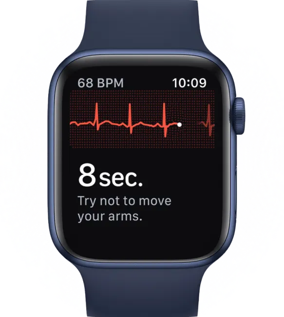
Many wearable devices, such as the Apple Watch, offer automated ECG readings. These provide a quick initial assessment, convenient for capturing data on the go. However, automated systems have their limitations. They excel at identifying straightforward normal sinus rhythms, but they may misinterpret more complex patterns or subtle variations. This can potentially lead to false positives or missed diagnoses.
This is where expert human analysis becomes invaluable. A certified cardiac technician (CCT) or a cardiologist brings years of training and experience to ECG interpretation. They can differentiate between benign variations and true abnormalities, taking into account individual factors such as age, gender, and medical history. These are often overlooked by automated systems. For example, a slightly prolonged QT interval might be normal for a woman but a cause for concern in a man. A computer might flag it as abnormal in both cases, creating unnecessary anxiety.
Technical Factors Affecting ECG Quality
Several technical factors can influence the accuracy of an ECG:
- Electrode Placement: Incorrect electrode placement can distort readings, leading to inaccurate interpretations.
- Patient Movement: Even small movements during the recording can create artifacts that resemble abnormalities.
- Electrical Interference: External electrical devices can interfere with the ECG signal, impacting the quality of the recording.
Careful preparation for your ECG, such as remaining still and following the technician's instructions, can minimize these issues.
Understanding the Limits and Benefits of a Single Lead ECG
A single lead ECG provides a snapshot of heart activity at a particular moment, which is beneficial for capturing episodes that might be missed during a doctor's visit. It is particularly useful for detecting certain heart rhythm irregularities that occur sporadically. However, it has its limitations and may not capture all types of heart issues, such as changes in the ST segment.
If you occasionally feel heart palpitations, a normal reading from a single lead ECG might not rule out an underlying problem. To thoroughly investigate, long-term monitoring or multiple ECG tests might be needed. It's important to consider ECG results together with your symptoms, medical history, and other tests for a full understanding.
Empower Your Heart Health with Qaly
Qaly offers advanced ECG interpretation services, connecting your at-home and wearable ECG recordings with certified cardiographic technicians. With human-reviewed analysis, you gain valuable, timely insights into your heart's electrical activity.
Not sure if your ECG is normal?. On the Qaly app, human experts will interpret your ECGs within minutes. Get started today.


.png)
.png)