

Key Takeaways

User Profile
Hello Heart Hero.
I know sometimes the healthcare world can feel confusing and you might have doubts. You are in the right place to learn what fluoroscopy is and how it can support your care.
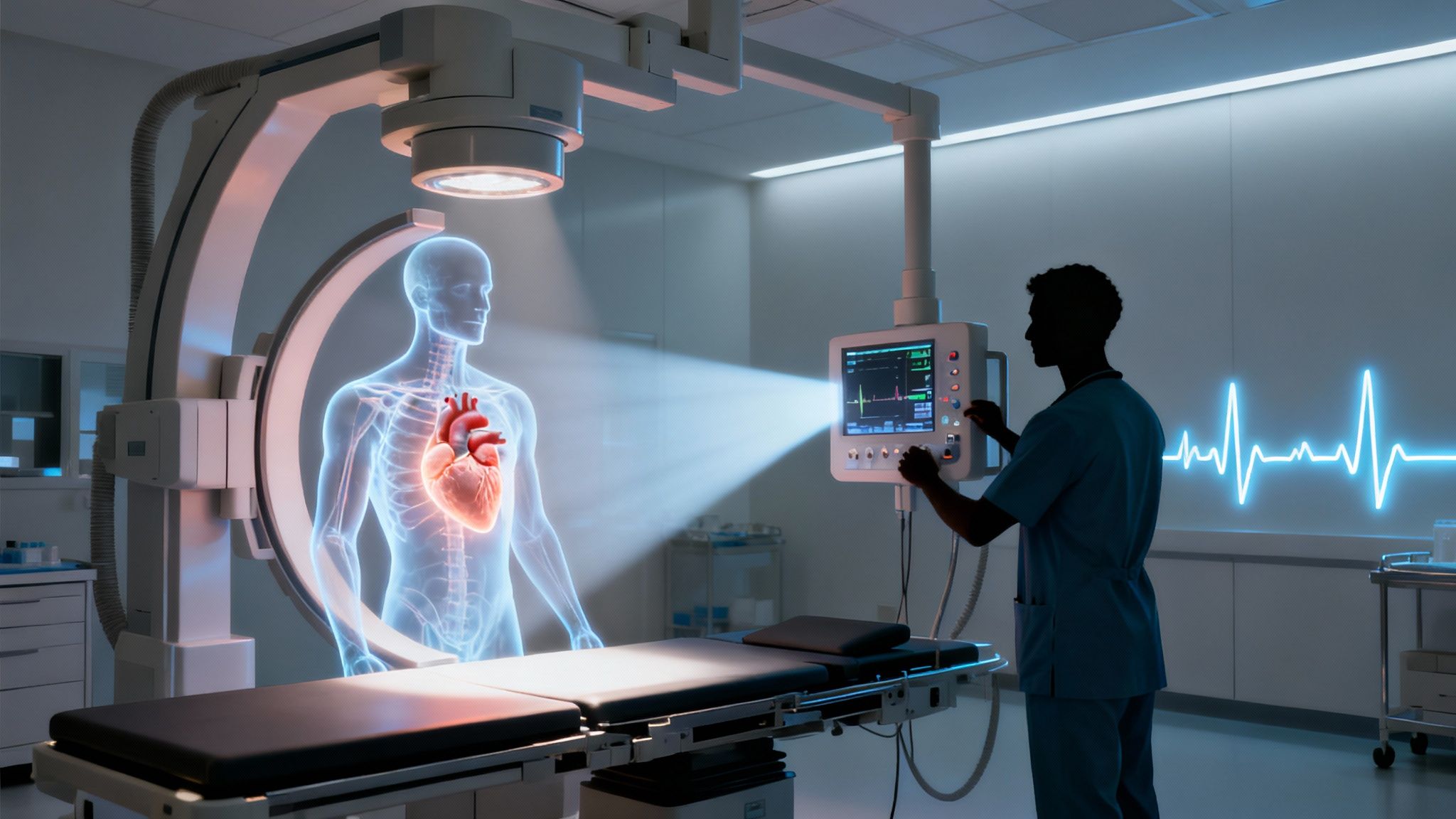
Fluoroscopy is like watching an X-ray movie playing live inside your body. Think of it as real-time imaging that captures your beating heart or contrast dye coursing through vessels.
Why does it matter? Live imaging guides treatment decisions on the spot - and when combined with wearable ECG data from Qaly, it delivers a fuller, clearer picture of your heart’s activity.
Quick Overview Of Fluoroscopy
At its core, fluoroscopy projects a continuous stream of X-ray images onto a monitor so you and your care team can watch internal structures move in real time.
Unlike a static snapshot, this approach reveals how contrast dye travels through arteries and exactly where catheters or stents are positioned.
Your cardiologist may recommend fluoroscopy for:
- Guiding stent or catheter placement during cardiac procedures
- Checking pacemaker or defibrillator lead positions in real time
- Assessing blood flow and valve function with contrast dye
- Supporting gastrointestinal and orthopedic studies if multi-system concerns arise
Pairing fluoroscopy with ECG insights from Qaly gives you a dynamic view of heart function. This synergy can spot subtle rhythm changes that a still ECG tracing alone might miss.
Key Takeaway Live imaging offers a dynamic look at heart motion to support timely treatment decisions.
Benefits Of Fluoroscopy
Seeing every movement as it happens means adjustments can be made on the spot.
This precision reduces guesswork when positioning devices or evaluating organ function.
And let’s be honest - knowing there’s a live view often brings extra confidence.
How It Works
An X-ray source emits beams through your body while a detector on the other side captures each fleeting moment.
Those raw signals are processed into a clear, continuous video on the display.
Understanding this process ahead of time can help calm nerves before your appointment.
In the next sections, you’ll learn about risks, safety precautions, and steps to take before, during, and after fluoroscopy so you can walk into your procedure fully prepared.
You’ll always feel supported every step of the way.
Understanding Key Concepts Of Fluoroscopy
Fluoroscopy transformed early X-ray snapshots into a live “movie” of your insides. Imagine watching your blood vessels and devices move in real time, rather than flipping through still photos.
- Real-Time X-Ray Stream shows anatomy in action
- Guides stent and catheter placement with precision
- Tracks the flow of contrast dye dynamically
- Complements ECG data for more complete heart insights
Fluoroscopy Compared To CT And MRI
While CT delivers static cross-section slices and MRI excels at soft-tissue detail, fluoroscopy streams continuous images. In practice, this means the cardiology team can steer wires and stents as if they’re playing a video game - making adjustments on the fly.
Continuous live imaging reduces uncertainty when positioning leads or checking implants.
Long before digital flat panels arrived, pioneers like Thomas Edison rigged fluorescent screens around 1900. By the 1930s, shoe-fitting fluoroscopes let shoppers glimpse their foot bones - sometimes exposing them to up to 13 roentgens in minutes. Historians estimate those retail demos packed a dose equal to 1,000 chest X-rays, leading to burns and long-term health risks. Discover more insights about early fluoroscopy history on PMC. That checkered past reminds us why modern safety measures matter just as much as the technology’s promise.
Why Motion Matters In Procedures
Think of fluoroscopy as a live video feed of your anatomy. That real-time view is invaluable during cardiac interventions like:
- Stent placement in blocked arteries
- Pacemaker lead positioning
- Catheter navigation in electrophysiology
Pairing these moving images with wearable ECG readings from Qaly means your care team can monitor both device position and heart rhythm simultaneously.
Modern fluoroscopy units use pulsed X-ray beams and digital subtraction to cut radiation by about 50%.
- Pulsed Fluoroscopy delivers radiation in short bursts
- Digital Subtraction enhances vessel visibility with minimal dose
If you’re curious about how ultrasound and fluoroscopy intersect, see What Is A Heart Echocardiogram.
By showing every twist and turn inside your vessels, fluoroscopy not only guides treatment but also gives you confidence in what’s happening behind the scenes. Now that you understand the origins, risks, and real-time power of this X-ray technique, you can face your next procedure informed and at ease.
How Fluoroscopy Works In Modern Practice
You might have spotted a fluoroscopy screen flashing in a clinic hallway. Behind that live X-ray movie is a pulsed beam scanning through your body, creating moving images in real time. Technicians steer these pulses - tweaking timing, frequency, and intensity - to get crisp pictures without overloading you with radiation.
For example, a pulsed beam mode at 7.5 to 15 Frames Per Second can cut your dose by half compared to older continuous X-ray methods. On top of that, modern flat-panel detectors have replaced bulky image intensifiers, boosting clarity while lowering skin-entrance dose by up to 70%.
- X-ray Tube Generates Pulses
- Body Absorbs and Transmits Signals
- Detector Captures Photons as Data
- Processor Converts Data Into Live Video
Key Insight Modern digital fluoroscopy platforms improve signal-to-noise ratios by 30-50%, making each session both safer and more precise.


Core Components And Their Roles
At its core, a flat-panel detector brightens faint X-ray signals much like turbo-charging a low-light photograph. Meanwhile, digital subtraction techniques remove background “noise,” isolating vessels and trimming contrast dye needs by 40% in many vascular studies.
This setup marks a sharp turn from the old days of fluorescent screens and bulky tubes. Today’s systems squeeze out better images at far lower doses Read more about dose and display advances.
This infographic walks you through each step - from the X-ray tube firing off pulses to the live video on the monitor - so you know exactly what’s happening.
Imaging Advances That Matter
- Digital Subtraction Angiography (DSA) subtracts a pre-contrast mask to spotlight blood flow, reducing lab doses by up to 50%.
- Instant Playback means clinicians can pause and zoom without interrupting the procedure.
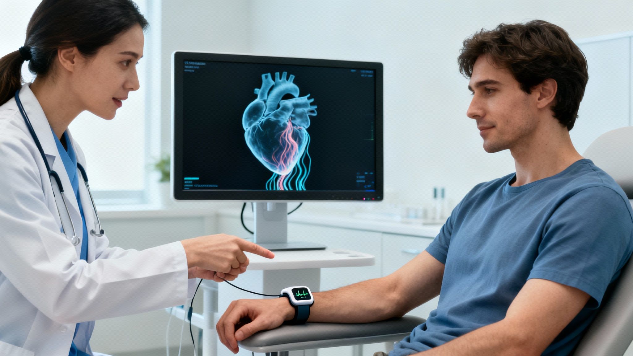
- Motion + Rhythm Insight pairs live fluoroscopy with ECG data from Qaly (Check out our guide on heart pacemaker operation), so your care team tracks structure and heartbeat in sync.
From hand-held screens in the early 1900s to pulsed digital systems by 2019, fluoroscopy technology has slashed doses by up to 70% while sharpening diagnostic accuracy Read the full evolution on FDA.
With this foundation, you’ll step into your next fluoroscopy with calm confidence.
Common Fluoroscopy Uses In Clinical Practice
Imagine you’re in the cath lab, watching a live X-ray image pop up next to your wearable ECG trace. Suddenly, you’re not just observing - you’re seeing your heart’s story unfold in real time.
Take stent placement, for instance. As the wire advances through a blocked artery, fluoroscopy highlights the dye flooding past the narrowing. Your cardiologist can tweak the stent on the spot, turning uncertainty into confidence.
In electrophysiology studies, catheters navigate blood vessels like expert spelunkers mapping a cave. Fluoroscopy follows every twist and turn, helping physicians zero in on the source of an arrhythmia while your ECG monitors each beat.
Then there’s pacemaker lead placement. Think of the lead as a cable anchoring a device inside your heart. Without fluoroscopy’s live feedback, those connections would be guesswork.
- Cardiac Catheterization Guides Stents And Balloons
- Electrophysiology Mapping Tracks Tiny Catheters
- Pacemaker And Defibrillator Lead Checks Verify Correct Position
Cardiac Procedure Examples
Consider a routine stent case. Under the X-ray beam, you see the vessel’s pinch point light up as contrast dye flows. At the same time, your Qaly ECG adds rhythm details - so the team watches both structural and electrical signals side by side.
If you want a deeper dive into rhythm studies, you’ll appreciate our guide on cardiac electrophysiology where imaging and ECG come together.
A look back at 400 bariatric patients after Roux-en-Y bypass exposed a 5.25% leak rate. Here, fluoroscopy caught subtle leaks that CT scans missed, steering treatment from diet tweaks to surgical repair. Learn more about these findings.
Interventional fluoroscopy sessions can exceed 100-500 mGy to skin, raising lifetime cancer risk, especially for younger women.
Other Clinical Applications
Fluoroscopy’s reach extends well beyond the heart.
- Gastrointestinal leaks and motility checks
- Spinal injections for back pain relief
- Guiding fracture repairs and prosthesis fitting
For example, in a follow-up swallow study, live X-rays revealed subtle esophageal spasms that an endoscope overlooked.
When your cardiologist requests both your wearable ECG data and a fluoroscopy exam, it isn’t redundancy - it’s two complementary lenses. Sharing Qaly data brings rhythm insights into the fluoroscopy suite, resulting in sharper diagnoses and treatment plans you can trust.
After your procedure, set aside time to review the findings and ECG trends with your cardiologist. It’s the final step in turning complex imaging into clear answers.
Risks And Safety Measures You Need To Know
For many, the word “radiation” triggers worry. But if you think of fluoroscopy like a live X-ray movie rather than a floodlight, it becomes easier to grasp why controlling exposure matters.
Depending on what’s happening under the hood, your treatment team might deliver:
- Gastrointestinal studies: 5–10 mGy per minute
- Cardiac catheterization: up to 50–100 mGy in longer cases
- Orthopedic injections: usually below 20 mGy
These figures can swing from just a few milligrays for a quick snapshot to hundreds for complex procedures. So dose management isn’t a nice-to-have - it’s essential.
Safety Protocols In Fluoroscopy
Your care team follows the ALARA principle - keeping radiation As Low As Reasonably Achievable. Diagnostic reference levels serve as guardrails, letting technologists tweak beam intensity on the fly. It’s a bit like adjusting camera brightness in a dim room: too bright washes out the detail, too dark hides what you need to see.
Shielding plays a starring role, too. Lead aprons and thyroid collars block stray rays, while beam collimation narrows the X-ray field to exactly where it’s needed. You can even ask for pulsed mode, which delivers X-rays in short bursts rather than a constant stream.
- Ask for a thyroid shield or lead apron
- Request tighter beam collimation to focus only on the target area
- Check if pulsed fluoroscopy or low-dose settings are available
When it comes to children and expecting moms, extra care is taken. Rapidly dividing tissues are more vulnerable, so doctors may postpone elective scans or opt for ultrasound or MRI instead.
“With skilled technologists and modern safety checks, fluoroscopy’s benefits almost always outweigh the risks.”
In the hands of an experienced team, fluoroscopy shines a light on hidden problems without undue exposure. Remember: your voice matters. If something feels off or you want more details, speak up.
Always loop in your cardiologist. Sharing your wearable ECG insights from Qaly alongside imaging data gives you the full picture of heart health. By asking the right questions and knowing what to expect, you stay one step ahead.
Empower yourself at every step - it’s your health, after all.
What To Expect During A Fluoroscopy Procedure
When you walk into the fluoroscopy suite, a friendly technologist greets you by name. They’ll show you to a quiet changing area, where you swap your clothes for a hospital gown - think of it as your backstage pass to the imaging show.
Next, you’ll remove any metal items - keys, watches, jewelry - to keep X-ray pictures crisp. A quick review of consent forms follows; this is your moment to ask questions and understand every step.
Your team then guides you onto the table, adjusting cushions under your back or legs so you stay relaxed. A monitor hums to life, displaying live X-ray images, and you’ll hear clear instructions on where to shift or hold still.
Key Steps During Your Exam
- Contrast Dye Injection: A brief warm flush spreading through your arm.
- Breathing and Positioning: Simple cues like “hold your breath” or “tilt slightly left” for the best shots.
- Continuous Monitoring: Technologists track your heart rhythm and image quality the entire time.
Most fluoroscopy sessions take 15–30 minutes, depending on what your doctor needs to see. It’s over before you know it.
“Step-by-step explanations help everyone stay calm,” one radiology tech shares.
When the imaging wraps up, you’ll slide off the table and sit briefly while staff check your vital signs. They’ll make sure you’re feeling fine before you head home.
Feel free to discuss any new ECG patterns from Qaly with your cardiologist. For more on cardiac procedures, check our guide on atrial fibrillation ablation.
Fluoroscopy Frequently Asked Questions
Many people wonder how long radiation lingers in their body. Think of it like sun-kissed skin - the glow fades in just a few hours. After your fluoroscopy exam, most of the X-ray dose dissipates from tissues quickly.
Eating and drinking before the procedure is usually fine unless your care team gives other instructions. To make your visit smoother:
- Ask About Fasting Guidelines to avoid discomfort.
- Inform Staff of any allergies or if you’re pregnant.
- Bring Your Qaly ECG Report so you can integrate live imaging with your heart rhythm data.
Fluoroscopy with ECG is like watching a movie and reading the script at the same time. You’ll see real-time X-ray images of your heart and vessels while your ECG trace scrolls alongside, giving clinicians a full picture of structure and rhythm.
When To Discuss Results With Your Cardiologist
If you notice new rhythm changes or any odd ECG patterns, share them right away. Your cardiologist uses both imaging findings and ECG trends to refine your treatment plan.
Combining live X-ray and ECG reduces uncertainty and speeds decisions.
Still have questions? Feel free to schedule a follow-up appointment and bring your curiosities with you.
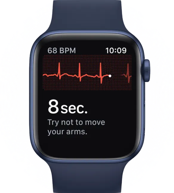
Have trouble interpreting your ECG? On the Qaly app, human experts will interpret your watch ECGs for arrhythmias and irregular heartbeats within minutes. Get started today.


.png)
.png)