


Key Takeaways

User Profile
A cardiac MRI is a safe, non-invasive imaging test that gives your doctor an incredibly detailed look at your heart. It uses a powerful magnet and radio waves, not radiation, to create precise pictures of your heart's structure and function, helping get to the bottom of symptoms like palpitations.
A Clearer Picture Of Your Heart Health
Hello, heart hero. We get it. Taking charge of your health, especially when you're tracking symptoms with a wearable device, can sometimes feel like trying to solve a puzzle without all the pieces. It's easy to feel a bit lost or anxious about what comes next in your healthcare journey, especially when you're feeling skeptical about the system. This guide is here to light up the path ahead.

Try to think of a cardiac MRI less as a daunting medical test and more as a high-definition 'map' of your heart. It’s a powerful tool that helps translate the electrical signals your watch picks up into a clear, physical picture of your heart's health. We’ll walk you through everything, breaking down the medical jargon into simple terms.
Why Your Heart Health Journey Matters
Feeling uncertain about what your symptoms mean is completely normal. The healthcare system can be tough to navigate, and it’s natural to want to find answers for yourself. By monitoring your health, you've already taken a huge and proactive step.
Our goal is to empower you with knowledge, connecting the dots between the data you see on your wrist every day and the advanced diagnostics that can provide definitive answers. A cardiac MRI is one of the best ways to achieve that clarity.
By looking directly at your heart's muscle, chambers, and valves, a cardiac MRI gives your doctor an unparalleled view of your cardiovascular health. It moves beyond just the electrical signals to show the physical engine that powers your body.
This advanced scan provides critical information that helps doctors diagnose and manage various heart conditions. It's especially useful when other tests, like an ECG or an echocardiogram, don't tell the whole story. You can learn more about the different forms of cardiac monitoring in our related guide.
Building Confidence in Your Care
Understanding what a cardiac MRI can reveal is the first step toward feeling more in control. It’s about turning that feeling of uncertainty into action and anxiety into answers.
This single scan can help explain a wide range of concerns, including:
- The underlying cause of arrhythmias or irregular heartbeats.
- The extent of damage after a heart attack.
- How effectively your heart is pumping blood.
- The presence of structural problems with your heart or major blood vessels.
Ultimately, a cardiac MRI is about providing peace of mind. Whether it confirms that everything is okay or pinpoints an issue that needs attention, the information it provides is invaluable. It equips you and your doctor with the insights needed to create a personalized and effective care plan, ensuring you're on the best possible path to long-term heart health.
What Is A Cardiac MRI And Why You Might Need One
Let’s talk about the cardiac MRI, a powerful scan that gives you and your doctor a remarkably clear window into your heart. When you’ve been carefully tracking your health and trying to get to the bottom of things, facing a new medical test can feel a little daunting. That's completely normal.
Think of it like trying to figure out what's wrong with a car engine just by listening to it. You might hear a clunk or a whir, but you can’t see the part that's causing the problem. A cardiac MRI is like popping the hood and getting a high-definition, slow-motion video of every single component as it works, matching a visual story to the sounds.
A Camera For Your Heart
At its core, a cardiac MRI uses a strong magnet and radio waves to create stunningly detailed, slice-by-slice pictures of your heart. It’s important to know that this scan does not use any radiation, setting it apart from other imaging like X-rays or CT scans.
This technology allows your doctor to see your heart’s physical structure, the chambers, the valves, and the major blood vessels connected to it. But it doesn't stop there. It also shows your heart’s function in real-time, revealing how well it’s pumping blood with every single beat. This one-two punch of structural and functional detail is what makes the MRI so incredibly useful.
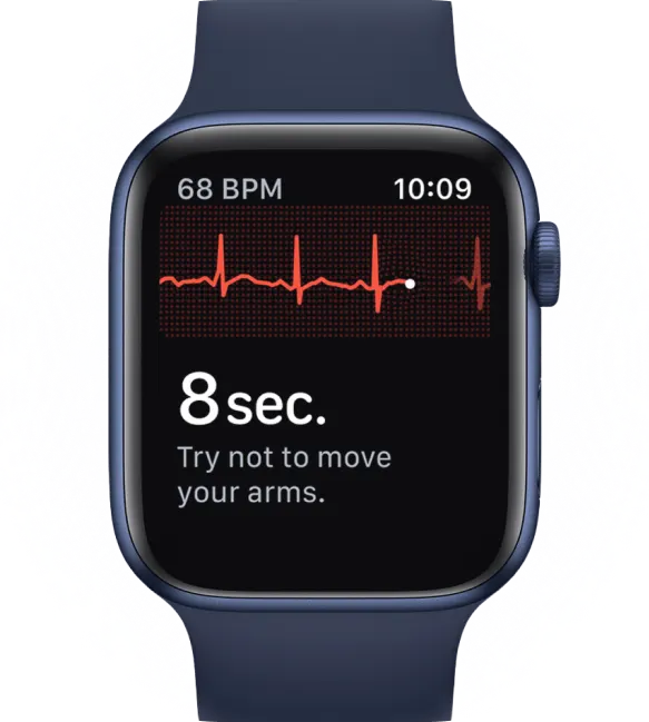
Think of it this way: if your wearable ECG is like a microphone picking up the rhythm of your heart, a cardiac MRI is like a high-definition video camera showing the physical condition of the orchestra producing that rhythm.
This dual insight is key to connecting the electrical data from your watch to the physical reality of what’s happening inside your chest. To see how other imaging tests stack up, check out our guide on the transthoracic echocardiogram, another common tool for visualizing the heart.
Why Your Doctor Might Recommend This Scan
A physician may suggest a cardiac MRI for a few important reasons, usually when other tests haven't provided a complete picture. It's often the final puzzle piece that explains the 'why' behind the symptoms you've been so carefully monitoring.
Here are some common situations where a cardiac MRI proves its worth:
- Investigating Arrhythmias: If you’ve been tracking palpitations or an irregular heartbeat in your app, an MRI can search for physical causes, like inflammation or scar tissue, that might be triggering those electrical glitches.
- Assessing Heart Muscle Health: Following an event like a heart attack, an MRI can map the exact location and extent of any damage. It's also excellent for spotting conditions like myocarditis, which is inflammation of the heart muscle.
- Examining Heart Chambers and Valves: The scan delivers crystal-clear images of your heart’s four chambers and valves, making it easy to spot leaks, blockages, or other structural issues impacting blood flow.
- Measuring Pumping Efficiency: It gives a precise measurement of how much blood your heart pushes out with each squeeze. This is a vital sign of overall heart function known as the ejection fraction.
This unmatched level of detail has made cardiac MRI a pillar of modern cardiology. Its use has grown since early studies confirmed its value. Today, with over 500,000 scans performed each year in the US, it’s a go-to diagnostic tool that helps reduce the need for more invasive procedures by up to 20-30% in certain cases. You can read more about the history of cardiac MRI’s growth and impact in cardiology.
For anyone looking for answers, this scan offers a definitive bridge between what you're tracking at home and a confident clinical diagnosis, giving you the clarity and peace of mind you need.
Your Step-by-Step Guide to the MRI Experience
Knowing what to expect can make a huge difference in easing any anxiety you might have about an upcoming scan. Medical tests can be stressful, we get it. So, let's walk through the entire cardiac MRI process from your perspective, so you feel prepared, comfortable, and in control.
The whole thing can be broken down into three simple phases: getting ready for the scan, the scan itself, and what happens after. Each part is pretty straightforward, and the medical team is there to help you the entire time.
Preparing for Your Appointment
Here’s the good news: getting ready for a cardiac MRI is usually a breeze. There are no complicated diets or fasting rules to worry about. The main thing is your safety, making sure the powerful magnet in the machine can do its work without any interference.
Before your appointment, someone from the imaging team will probably call you to run through a safety checklist. This is the perfect time to ask any questions you have. They'll ask about any metal you might have in your body, which is the most critical part of the prep.
Here’s a quick rundown for the day of your scan:
- Dress for Comfort: Go for loose-fitting clothes without any metal. This means no zippers, snaps, or even metallic threads. Think sweatpants and a simple t-shirt. Most places will give you a hospital gown to change into anyway.
- Leave the Metal Behind: Take off all jewelry, watches, hearing aids, and piercings before you even leave the house. You’ll have to remove them before the scan, so it's just easier to leave them safe at home.
- Confirm Your Implants: If you have a pacemaker, defibrillator, cochlear implant, or any other medical device, make sure you've talked it over with both your doctor and the MRI staff. Many modern devices are MRI-safe, but the team needs to know the exact make and model to be sure.
During the MRI Scan Itself
Once you've changed and gone through a final safety check, the MRI technologist will bring you into the scanner room. This person is your guide for the entire test. They're highly trained professionals whose main job is to keep you as comfortable as possible.
You'll lie down on a padded table that slides gently into the center of the MRI machine, which looks like a big, open-ended tunnel. The tech will put some small ECG patches on your chest to keep an eye on your heart rate and might give you headphones to muffle the noise and talk to you during the scan.
You can talk to the technologist at any time through an intercom. You're never alone in there, and you can always ask for help or a break if you need one.
The machine makes a series of loud thumping, buzzing, and clicking sounds as it takes the pictures. This is completely normal. The headphones often play music, or you can use earplugs to soften the noise. The most important thing for you to do is to lie as still as you can so the images come out clear. The whole scan usually takes between 45 to 90 minutes.
As you can see, an MRI is often a key step when your symptoms and initial tests suggest that a closer look is needed.
After the Scan Is Complete
Once the last picture is taken, the table will slide back out, and the tech will help you up. And that's it, you're done! There's no recovery time needed at all. You can change back into your clothes and get right back to your day.
You won’t get your results immediately. A specialized radiologist or cardiologist has to carefully look at the hundreds of detailed images taken during your scan. They'll write up a full report and send it to the doctor who ordered the test, which typically takes a few days. Your doctor will then schedule a follow-up with you to go over what they found.
This technology has come a long way. The journey of cardiac MRI started changing heart diagnostics back in the early 1980s, marking a huge shift from basic X-rays to detailed images that could see inside a beating heart without any surgery. Pioneers captured the first moving images of infant hearts in 1981 and 1983, dramatically cutting scan times and making the process much easier for patients. You can read more about the pioneering history of cardiac MRI imaging to see how far we've come.


What A Cardiac MRI Reveals About Your Heart
A cardiac MRI gives your doctor an incredible look inside your heart, showing important details that other tests simply can't capture. Instead of just getting a list of technical findings, let’s explore what the results from this powerful scan actually mean for your health and your peace of mind.
Think of it as the ultimate blueprint of your heart. It doesn't just show one aspect; it examines the entire system, from the muscle that powers every beat to the valves that control blood flow. This detailed view helps connect the dots between the symptoms you feel and the physical condition of your heart.
Peering Inside The Heart Muscle
One of the most valuable things a cardiac MRI does is assess the health of your heart muscle, also known as the myocardium. It can spot issues like inflammation (myocarditis) or scarring from past, sometimes silent, heart events.
Imagine your heart muscle as a strong, flexible rubber tire. A scar on that muscle is like a stiff, inflexible patch on the tire. This damaged area can't contract and relax properly, which can affect how well your heart pumps blood. An MRI can pinpoint the exact location and size of these scars, helping explain symptoms like fatigue or shortness of breath.
Checking The Heart's Plumbing and Valves
Beyond the muscle, the scan provides a crystal-clear view of your heart’s four chambers and the valves that act as one-way doors between them. It checks to see if these valves are opening and closing correctly.
- Leaky Valves (Regurgitation): The MRI can show if a valve isn’t closing tightly, allowing blood to flow backward.
- Narrowed Valves (Stenosis): It can also detect if a valve has become stiff or narrowed, forcing the heart to work harder to push blood through.
These valve issues can be a source of irregular heart rhythms or feelings of lightheadedness, and an MRI can measure their severity with incredible accuracy.
Measuring Function and Blood Flow
A cardiac MRI doesn't just give you a static picture; it creates a dynamic movie of your beating heart. This allows doctors to measure exactly how efficiently your heart is pumping blood throughout your body with each beat. This measurement is called the ejection fraction.
A healthy ejection fraction is a key indicator of good heart function. If it’s low, it tells your doctor that your heart is struggling, which helps guide treatment decisions. The scan also tracks blood flow through the major vessels, looking for blockages or structural problems you may have been born with.
A cardiac MRI provides a complete picture, showing not just the heart’s structure but also its real-time performance. It’s like getting both the architect's blueprints and a live video feed of how the building is functioning day to day.
The Power of Gated Imaging
Capturing a clear image of a constantly moving organ is a huge technical challenge. Early on, this was a major hurdle. The breakthrough came with ECG-gated cardiac MRI, a technique introduced in the 1980s that synchronized the imaging with the heart's own rhythm. By timing the pictures to a specific point in the heartbeat (the R-wave on an ECG), the MRI could effectively "freeze" the motion.
This advance was a game-changer, allowing for precise measurements of chamber volumes and function, often with an accuracy within 5% of more invasive tests. By the 1990s, this technology had become so refined that it could measure heart volumes with an accuracy of ±10 ml, far surpassing the limits of other imaging methods at the time. Discover more insights about the history of magnetic resonance imaging at rbht.nhs.uk.
By revealing both the physical structure and the functional performance, a cardiac MRI provides an all-in-one assessment of your heart's overall condition, giving you and your doctor the clear answers needed to move forward confidently.
Connecting Wearable ECG Data With MRI Results
This is where your proactive health tracking really pays off. It is about connecting the dots between what you feel every day and what your doctor can now see in incredible detail. You’ve been gathering valuable data with your wearable ECG, and your cardiac MRI has provided a precise structural map of your heart. Now it’s time to bring these two powerful stories together.
This process can be incredibly empowering. Instead of just receiving test results, you get to be an active partner in your own healthcare conversation. Let’s walk through exactly how to do this, making sure your insights are heard and understood.
Organizing Your ECG Data for a Productive Conversation
Walking into your doctor's office with a mess of unorganized data can be overwhelming for everyone. The goal is to present your information in a clear, organized way that tells a compelling story. Think of yourself as a detective laying out the key evidence.
Before your follow-up appointment, spend some time reviewing your ECG reports. You’re looking for patterns or specific events that jump out.
- Identify Key Events: Pinpoint the dates and times of significant symptoms, like frequent palpitations, a racing heart, or moments you felt dizzy or lightheaded.
- Highlight Trends: Have your symptoms become more frequent? Do they tend to happen at certain times of the day, or after specific triggers like exercise or caffeine?
- Prepare Your Reports: An app can help you organize and print your ECGs. Having a physical or digital folder ready makes the whole conversation flow much better.
As you get your data together, don't forget the importance of privacy. Using tools for secure document sharing ensures your sensitive health information stays confidential when you send it to your healthcare providers.
Framing Questions That Connect Your Data to Your Diagnosis
Once your data is organized, you can start framing smart, specific questions that link your lived experience to the clinical findings from the MRI. This simple step elevates the conversation from a one-way report into a collaborative analysis of your heart health.
Instead of a general question like, "So, is my MRI normal?" you can be much more direct and insightful. Your goal is to help your doctor see the connection between the electrical "blips" you've recorded and the physical structure of your heart.
By presenting your ECG data alongside your MRI results, you're not just asking what the scan shows; you're asking what the scan means for you and the symptoms you're experiencing.
Examples of Powerful, Data-Driven Questions
Here are a few examples of how you can frame your questions to spark a more meaningful discussion with your doctor:
- Connecting Palpitations to a Structural Finding: "I've recorded frequent palpitations on my ECGs, especially in the evenings. My MRI report mentions a mild mitral valve leak. Could these two things be related? Is it possible the leak is causing the palpitations I'm feeling?"
- Linking Fatigue to Pumping Function: "My wearable has been tracking a consistently high resting heart rate, and I've been feeling really fatigued. The cardiac MRI showed my ejection fraction is 50%. How does this number connect to the fatigue and high heart rate I've been tracking?"
- Questioning Scar Tissue and Arrhythmias: "The MRI found a small area of scarring on my left ventricle. I captured this ECG showing an irregular rhythm during a workout last week. Is it possible that the scarred tissue could be the source of this arrhythmia?"
This proactive approach does more than just get you answers. It builds trust and shows your doctor that you're an informed, engaged partner in managing your health. It ensures your care plan isn't just based on a single test, but on a complete picture of your unique health journey.
Common Questions About Cardiac MRI Scans
Going in for a medical scan can feel a little intimidating, and it’s completely normal to have questions. Getting clear answers is the best way to quiet any anxiety and feel more in control of your health. Let’s walk through some of the most common questions people have about the mri cardiac experience.
Is A Cardiac MRI Safe And Does It Use Radiation
This is one of the first questions on everyone's mind, and the answer is reassuring. A cardiac MRI is considered extremely safe, and here’s the most important part: it does not use any ionizing radiation. Unlike X-rays or CT scans, this technology creates incredibly detailed images using a powerful magnet and radio waves.
The primary safety concern revolves around metal. Because the machine is a giant magnet, the medical team will thoroughly screen you to make sure there’s no metal inside your body that could cause a problem. This includes things like pacemakers, defibrillators, or even older surgical clips. Many modern medical implants are now designed to be MRI-safe, but it's absolutely critical to tell the MRI team about every single implant you have so they can ensure your complete safety.
Will I Need An Injection For My Cardiac MRI
Often, yes, an injection is part of the scan. Many cardiac MRI exams use a contrast agent called gadolinium, which is given through a small IV line in your arm.
Think of it like adding a drop of food coloring to a glass of water to see the currents inside. This contrast agent travels through your bloodstream and highlights different areas of your heart muscle. This makes it much easier for the radiologist to spot things like scarring, inflammation, or problems with blood flow. The agent is generally considered very safe and is well-tolerated by most people.
As a standard precaution, the team will ask you about your kidney function, since your kidneys are responsible for filtering the contrast out of your system. If you have known kidney issues or any allergies, be sure to bring them up with your doctor and the MRI staff beforehand.
How Should I Prepare For My Cardiac MRI Appointment
The good news is that preparation is usually pretty straightforward. It's all about making sure you're safe and comfortable. You can typically eat and drink as you normally would, though some centers might ask you to skip caffeine for a few hours just because it can speed up your heart rate.
The most important preparation step is removing all metal from your body. This is a strict safety rule.
- Clothing: Plan on wearing comfortable clothes with no metal. Think sweatpants and a plain t-shirt.
- Accessories: Leave all jewelry, watches, and hearing aids at home or in a locker. You’ll have to take them off anyway.
- Questionnaire: You'll fill out a detailed form about your medical history, specifically asking about any past surgeries or implants.
If you’re worried about feeling claustrophobic in the scanner, a very common concern, let your doctor know ahead of time. They can sometimes prescribe a mild sedative to help you relax. Don't hesitate to share any concerns with the MRI team; they're there to make the experience as smooth as possible for you.
What Does It Mean If My Cardiac MRI Mentions Troponin Levels
Sometimes, your doctor will look at your MRI results alongside other tests, like a blood test that measures your troponin levels. Troponin is a protein that your heart muscle releases into the blood when it's been damaged, like during a heart attack.
The two tests work hand-in-hand. The blood test showing high troponin tells your doctor that damage has occurred, and the cardiac MRI can then show the physical evidence of that damage, like a scar on the heart muscle. It can pinpoint exactly where the injury happened and how big it is. It connects the "what" from the blood test to the "where" and "how much" on the image. To learn more, check out our guide on what troponin levels tell you about your heart.
By understanding these key parts of the process, you can walk into your appointment feeling prepared and empowered. The whole point of an mri cardiac scan is to get clear answers, and knowing what to expect is the first step.
At Qaly, we believe that understanding your heart shouldn't be confusing. Our expert-reviewed ECG analysis for your smartwatch helps you make sense of your symptoms, track your heart's rhythm, and have more productive conversations with your doctor


.png)
.png)