

Key Takeaways

User Profile
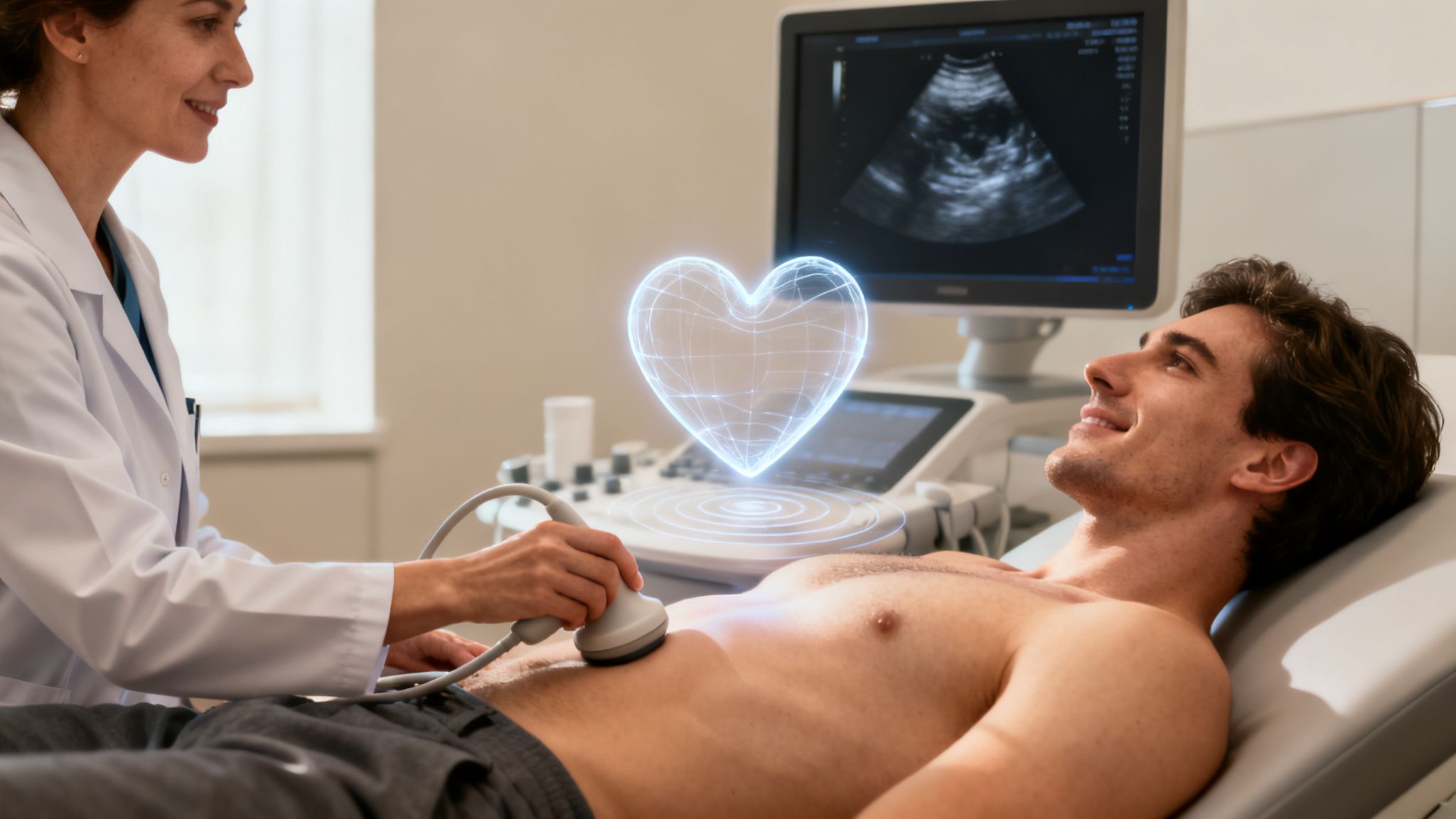
Hello, heart hero. We get it. Hearing you need another heart test can be stressful, especially when you're already trying to make sense of palpitations or strange rhythms from your wearable device. It's easy to feel a little skeptical, wondering what this new test will actually show. This guide is here to walk you through the transthoracic echocardiogram (TTE), explaining exactly what it is with clarity and empathy.
A Clearer Picture of Your Heart Health
Think of a transthoracic echocardiogram as a detailed ultrasound movie of your heart. While the ECG on your watch is brilliant at tracking your heart's electrical rhythm, the TTE shows its physical structure and function. It gives us a live view of your heart's pumping strength, a look at how your valves are working, and the exact size of its chambers.
This test is a cornerstone of heart care for a simple reason: it's safe, painless, and incredibly insightful. It uses sound waves, just like the ultrasounds used during pregnancy, to create a moving, real-time image of your heart. There is zero radiation involved, making it one of the most common and trusted tools a cardiologist has.
Why This Test Is So Common
The transthoracic echocardiogram is the most frequently performed type of heart ultrasound, and for good reason. It’s accessible and cost-effective, making it the first choice for doctors who need a comprehensive look at the heart's mechanical function without resorting to invasive procedures.
Its widespread use is reflected in its market dominance. The global echocardiography market is projected to experience significant growth by 2032. This expansion is driven by the increasing demand for reliable heart diagnostics, particularly as more individuals manage heart conditions. For perspective, in 2021 alone, over 7.6 million people in the UK were living with heart or circulatory diseases, highlighting the global need for effective tools like the TTE. You can explore more of these trends in the full report.
What Makes a Transthoracic Echocardiogram Different
It helps to understand where this specific test fits into your overall care. You’ve probably heard of other heart tests, but the TTE offers a unique and foundational perspective.
- Electrical vs. Mechanical: Your smartwatch ECG measures the electrical signals that tell your heart to beat. A TTE, on the other hand, shows the mechanical response to those signals, how strongly the heart muscle actually contracts.
- Non-Invasive Nature: Unlike some detailed imaging tests, the TTE is completely external. A small wand, called a transducer, is simply moved across your chest with some cool gel.
- Real-Time Imaging: The test provides a live video, allowing the sonographer to see exactly how your heart valves open and close and how blood moves through the chambers with each and every beat.
This procedure is about getting answers. It’s a way to move past the uncertainty that comes with unusual symptoms or confusing ECG readings from a wearable, giving you and your doctor a solid, structural foundation for your diagnosis.
Understanding what a transthoracic echocardiogram involves can help you feel more in control and less anxious about your appointment. It’s a proactive step toward a complete picture of your heart's health, empowering both you and your doctor with the information needed to make the best decisions for your care.
Why Your Doctor Recommended This Heart Ultrasound
If your doctor just suggested a transthoracic echocardiogram, you might be feeling a little worried or even confused. After all, you've been keeping a close eye on your heart with your wearable device, so why another test?
It's a fair question. But think of this recommendation as a really positive step toward getting the full picture of your heart's health, not a reason to panic. Your doctor is following established Evidence Based Practice Guidelines which help them make the best decisions for your care based on your specific symptoms and the data you've collected.
Connecting Your ECG Data to Your Heart's Structure
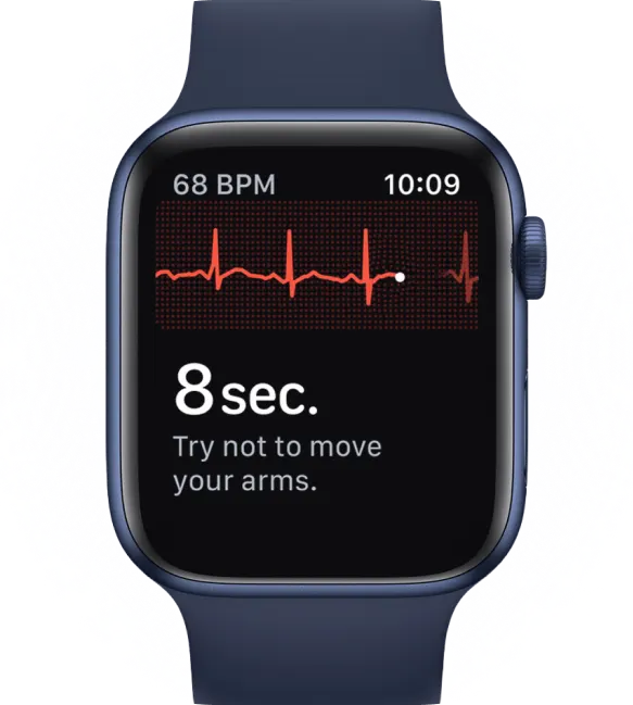
Your at-home ECG is an amazing tool. When a service like Qaly flags an arrhythmia like atrial fibrillation (AFib), frequent PVCs, or SVT, it's giving you a critical piece of the puzzle, the electrical piece.
But your ECG can't see the why behind that electrical pattern. It's a bit like hearing a weird noise coming from your car's engine. You know something's off, but you need a mechanic to pop the hood and see what's actually going on inside. The transthoracic echocardiogram is that look under the hood for your heart.
This heart ultrasound gives us direct answers to physical questions an ECG just can't tackle:
- How are the valves working? Are they opening and closing correctly, or is one a bit leaky?
- Is the heart muscle too thick? We can check for a condition called hypertrophy, where a wall of the heart has thickened.
- How strong is the pump? A crucial measurement called ejection fraction tells us exactly how efficiently your heart is pumping blood.
- Are the chambers the right size? Sometimes, if the heart has been working too hard, its chambers can become enlarged.
A transthoracic echocardiogram helps connect the dots between the symptoms you feel, the rhythm data from your device, and the actual physical health of your heart. It’s the bridge between the electrical signals and the mechanical function.
A Frontline Tool for Modern Heart Care
In cardiology today, the TTE has become absolutely essential, especially with heart health being a growing concern for millions. It's a go-to diagnostic tool for good reason.
With cardiovascular disease on the rise, over 7 million TTEs are performed in North America alone each year. Because it’s non-invasive, radiation-free, and relatively low-cost, it's the perfect first step for getting clear, actionable answers about your heart's structure and function.
For example, if your ECG shows a pattern that might be related to heart failure, the echocardiogram is what gives us the definitive look at your heart's pumping strength. If you’re curious about what those signs look like, you can learn more about understanding heart failure on your ECG in our detailed guide.
Ultimately, this test isn't just another appointment. It's the best way to get certainty, validate the data from your wearable, and give your doctor the visual evidence they need to create a precise, personalized plan just for you.
What Happens During the Echocardiogram Procedure
We know that walking into any medical appointment, especially a new one, can be a little nerve-wracking. That’s completely normal. Our goal here is to pull back the curtain on the transthoracic echocardiogram so you know exactly what to expect, step by step. When you feel prepared, you feel more in control.
Often, the journey to a TTE begins long before you even schedule the test. It might start with an alert from your wearable device, a conversation with your doctor about your symptoms, and then a recommendation for this detailed look at your heart.
This simple flow shows how your proactive health monitoring connects directly to getting a deeper, structural view of your heart.
The Calm and Quiet Setting
First, let’s set the scene. You'll be led into a private exam room, which is usually dimly lit. This isn't to create a gloomy mood; it's purely practical. The low light helps the sonographer, the highly trained specialist performing your test, see the ultrasound images on the monitor with perfect clarity.
You’ll be asked to undress from the waist up and given a hospital gown to wear for privacy and comfort. Then, you'll get settled on a comfortable examination table, usually lying on your back or your left side.
The Procedure: A Step-By-Step Walkthrough
Once you’re comfortable, the sonographer will start the test. The whole process is designed to be as easy and straightforward as possible. Here’s what you can expect:
- Placing the Electrodes: The sonographer will place a few small, sticky patches called electrodes on your chest. These are connected to an electrocardiograph (ECG) machine that tracks your heart’s electrical rhythm during the test. This helps sync the ultrasound pictures with your actual heartbeat.
- Applying the Gel: Next, a clear, water-based gel is applied to your chest. It might feel a bit cool and wet, but it's completely harmless. This gel is crucial because it eliminates any air between the probe and your skin, allowing the sound waves to travel cleanly to your heart.
- Using the Transducer: The main part of the test uses a small, handheld device called a transducer. The sonographer will press this wand gently against your skin and move it around your chest. You’ll feel some light pressure, but it's completely painless. The transducer sends out harmless sound waves and picks up the echoes that bounce back from your heart, creating the live images you see on the screen.
- Capturing the Best Views: To get the clearest pictures, the sonographer might ask you to do a few simple things. This could mean changing positions, like rolling onto your side, or briefly holding your breath. These small adjustments help move your lungs out of the way, giving the transducer an unobstructed view of your heart.
The entire transthoracic echocardiogram is non-invasive. There are no needles, no radiation, and no pain. It’s simply a way of using sound to look inside and see how your heart is doing its incredible job, beat by beat.
Typically, the entire procedure takes between 30 to 60 minutes. Throughout the test, the sonographer is focused on capturing high-quality images while making sure you’re comfortable. If you have any questions or feel uneasy at any point, don't hesitate to let them know.
Understanding the whole process can make the experience feel much more relaxed. If you're looking for a broader overview, you might find our guide that asks the simple question, what is a heart echocardiogram?, helpful. This simple, reassuring test is a powerful way to gain peace of mind and get the detailed answers you and your doctor need.


How to Understand Your Echocardiogram Results
Getting a medical report can feel like trying to read another language. It’s usually packed with technical terms that can leave you feeling confused and maybe a little anxious. After all, this is your health, and you deserve to understand what the results of your transthoracic echocardiogram actually mean.
Think of us as your translator. By getting a handle on a few key concepts, you can go into your follow-up appointment feeling confident and ready to be an active part of the conversation about your health. Let's break down the most common findings you'll see.
Measuring Your Heart’s Pumping Power
One of the most important numbers on your report is the Ejection Fraction (EF). It sounds complex, but the idea behind it is pretty simple. It’s basically a measurement of your heart's raw pumping power.
Your heart fills with blood and then squeezes to pump that blood out to the rest of your body. The ejection fraction is the percentage of blood that gets pushed out of the left ventricle, your heart's main pumping chamber, with each beat.
Here is a simple breakdown of what the numbers mean. An EF between 50% and 70% is typically considered normal. This shows your heart is pumping a healthy volume of blood with every beat. An EF from 41% to 49% might suggest that your heart's pumping ability is a bit weaker than it should be. Finally, an EF of 40% or less is a sign that the heart muscle isn't pumping effectively, which can be an indicator of heart failure.
This single number gives your doctor a powerful snapshot of how well your heart is doing its job.
Checking the Doors of Your Heart
Your heart has four valves that work like one-way doors, making sure blood flows in the right direction and doesn't back up. A transthoracic echocardiogram gives your doctor a crystal-clear look at how well these doors are opening and closing.
Two common valve problems that might show up on your report are:
- Valvular Stenosis: This is when a valve gets stiff and doesn't open all the way. Imagine a door that’s stuck and can only open a crack. Your heart has to work much harder to force blood through that narrowed opening.
- Valvular Regurgitation: This happens when a valve doesn’t seal tightly when it closes, letting some blood leak backward. Think of a door with a faulty seal that lets a draft seep back in. This leak forces your heart to work overtime.
Your report will point out which valve is affected and grade the problem as mild, moderate, or severe.
Your echocardiogram report isn't a final judgment; it's a detailed map. It shows your doctor the exact areas that need attention and helps them chart the best course forward for your health, turning you from a worried patient into an informed advocate.
Sizing Up the Rooms of Your Heart
Just like a house has different rooms, your heart has four chambers. The echo measures their size and wall thickness to see if they're under any strain.
- Chamber Enlargement: If one of the chambers is larger than normal, it could be a sign that it’s been working under pressure. For instance, if your smartwatch flagged atrial fibrillation, your echo might reveal an enlarged left atrium.
- Hypertrophy: This is the medical term for a thickened heart muscle wall. It's often the heart's way of adapting to high blood pressure or a stiff valve, the muscle bulks up to handle the extra workload.
Modern imaging is making these measurements incredibly precise. Advanced tools like 3D transthoracic echocardiography are changing the game in cardiac diagnostics, with 75% of surveyed centers now using the technology. For patients whose irregular rhythms were first picked up on a wearable device, these detailed images are crucial. You can learn more about the impact of these advanced techniques in cardiac care.
Understanding these core concepts from your transthoracic echocardiogram report is empowering. It helps you ask smarter questions and really grasp the "why" behind any of your doctor's recommendations. Sometimes, heart muscle strain can also be seen in blood tests. You can read our guide on what your troponin levels mean for your heart to learn more.
Connecting TTE Insights with Your Wearable ECG Data
This is where the two sides of your heart's story come together. You've been diligently tracking your heart's rhythm with your watch, and now you have the detailed, visual results from a transthoracic echocardiogram. So, how do these two powerful pieces of information connect?
This is the moment you go from simply collecting data to truly understanding what's going on. Your wearable ECG is like a personal rhythm detective, fantastic at catching the electrical patterns of your heart. The TTE provides the crucial structural context, showing the physical reason, or lack thereof, behind those electrical signals.
From Electrical Signals to Structural Certainty
Think of it this way: your ECG data shows the notes in a song, while your TTE lets you see the actual instrument playing them. One tells you what is being played; the other shows you the condition of the instrument itself. It's an incredibly powerful combination for getting clear answers.
For example, let's say your watch has been flagging frequent premature ventricular contractions (PVCs). That can be unsettling, but a TTE can offer profound peace of mind. If the echo shows that your heart's structure, valves, and pumping function are all completely normal, those PVCs are very often benign. Now you have solid proof that there isn't an underlying structural problem causing them.
By pairing your daily rhythm monitoring with a detailed structural exam like the transthoracic echocardiogram, you and your doctor get the most complete and actionable view of your heart, leading to a truly personalized care plan.
Creating a Complete Picture for Your Doctor
When you bring both sets of data to your cardiologist, you're giving them a much richer story than either test could tell on its own. It’s the difference between a single snapshot and a full documentary of your heart's health.
Here’s how they complement each other for common conditions:
- For Atrial Fibrillation (AFib): Your watch ECG is great at detecting the irregular rhythm itself. But the TTE measures the size of your left atrium, an important detail, as an enlarged atrium can influence how AFib is managed.
- For Unexplained Shortness of Breath: You feel breathless, but your ECGs look normal. A TTE can check for problems with your heart's pumping strength (ejection fraction) or valve function that could be the hidden cause.
- For Heart Palpitations: Your ECG confirms when you feel palpitations. The TTE helps figure out why by checking for structural issues like a mildly leaky valve (mitral valve prolapse) that might be contributing to the sensation.
How to Share Your Data Effectively
Bringing this information to your doctor is a key part of advocating for your health. Don't just say you have some ECGs on your phone; show up prepared with an organized report.
Apps like Qaly let you compile your ECG readings into a clear, shareable PDF report that your doctor can review right alongside your TTE results. This helps you have a more productive, evidence-based conversation. For more on getting the most out of your device, check out our cardiologist's guide to the smartwatch ECG for practical tips.
Ultimately, by pairing your wearable data with the clinical insights from a transthoracic echocardiogram, you shift from being a passive patient to an informed partner in your own healthcare. You're no longer just listening to what the system tells you; you're actively contributing the detailed, personal data needed for the best possible outcome.
Frequently Asked Questions About the Procedure
It's completely normal to have a few questions running through your mind before any medical test. Knowing what to expect can make all the difference. Let's walk through some of the most common questions people have about a transthoracic echocardiogram.
Our goal is to clear up any uncertainty so you can walk into your appointment feeling confident and at ease.
Is a Transthoracic Echocardiogram Painful or Dangerous?
Let's get this one out of the way first: absolutely not. It’s one of the safest and most routine ways we have to look at the heart.
A transthoracic echocardiogram uses sound waves, not radiation, so it's as safe as the ultrasounds used during pregnancy. The only things you'll feel are the cool ultrasound gel on your skin and the gentle pressure from the transducer as the sonographer moves it across your chest to get the best pictures.
You can relax knowing this is a completely painless and non-invasive test. There are no needles and no discomfort involved. It's simply about capturing images of your heart from the outside.
How Long Does It Take to Get My Echocardiogram Results?
This can vary a bit from place to place. The sonographer captures all the live "video" of your heart during the test, but the next crucial step is for a cardiologist to meticulously review those images and write up a formal report.
In many clinics, this report is ready for your doctor within a few business days. The best thing to do is ask the sonographer or front desk staff about their typical turnaround time before you leave. And make sure you have a follow-up appointment scheduled with your own doctor to go over the findings in detail.
Do I Need Any Special Preparation for the Test?
One of the great things about this test is how simple the preparation is. For the most part, you don't need to do anything special at all.
- Eating and Drinking: You can eat, drink, and go about your day as you normally would. No fasting required.
- Medications: Take all your regular medications as scheduled unless your doctor has given you specific instructions to do otherwise.
- Clothing: A comfortable, two-piece outfit is a good idea. You’ll be asked to undress from the waist up and wear a gown, so separate tops and bottoms just make things a little easier.
That's it. The easy prep is just another reflection of how straightforward and non-invasive the test is.
Take control of your heart's story. With Qaly, you can get your wearable ECGs analyzed by certified experts in minutes, giving you the clear, reliable data you need to have informed conversations with your doctor.


.png)
.png)