


Key Takeaways

User Profile
Hello Heart Hero. You're folding laundry, loading the dishwasher, or standing in the kitchen when a sudden chest pain shoots into your back and your smartwatch ECG becomes the first thing you reach for. That moment can feel isolating, especially if you've already had to fight to be taken seriously by the healthcare system. Aortic dissection ECG questions live in that space between fear and uncertainty, where a wearable can help you notice danger, but it can't give you the final answer.
The hard part is that a dissection can look quiet on an ECG, or it can look a lot like something else. A normal tracing may reassure you in the wrong way, while an abnormal one may push attention toward a heart attack when the true problem is a torn aorta. The safest path is to understand what your wearable can show, what it can't, and when the next step is urgent imaging rather than more waiting.
Introduction
You might already know the feeling of checking your watch and hoping for a clear, simple result. Then you see a tracing that looks off, or it looks fine while your body feels anything but fine. That mismatch is exactly why aortic dissection ECG knowledge matters for wearable users.
Aortic dissection is uncommon, but it carries high stakes. It tends to show up as a small share of emergency cases, and it more often affects adults in midlife than younger people. A smartwatch ECG can still be useful in that moment, because it may raise concern, but it cannot safely rule out aortic catastrophe on its own.
If symptoms are sudden, severe, or spreading, the goal is not to keep refreshing the tracing. The goal is to get the right kind of urgent care fast.
Understanding ECG Role in Aortic Dissection
A wearable ECG records the heart's electrical activity, not the aorta itself. When the aorta tears, the problem is structural, but the tracing can still shift because blood flow, coronary perfusion, and heart strain may be affected. That is why the ECG can act like a dashboard warning light. It may point to trouble, even when the source of the trouble sits outside the heart.
What the ECG can and can't tell you
The main point is straightforward. A normal ECG does not exclude dissection. The ECG works best as a rapid triage tool, not a rule-out test. It can add concern, but it cannot close the case on its own.
That matters because dissection is uncommon, so chest pain is often first linked to more familiar causes. In acute care, it appears in only a small slice of patients with chest, back, or abdominal pain, yet the stakes are far higher than a routine palpitations check. The ECG helps sort risk and guide urgency, but it does not replace imaging.
A green light on a wearable can still be misleading if the person has sudden tearing pain, faintness, or other warning symptoms. A yellow or red tracing does not identify the exact problem source, it only shows that something deserves attention. For wearable ECG users, the safest reading is the one that keeps the whole clinical picture in view, not the watch alone.
For a closer look at how ST changes are tracked and interpreted in ECG monitoring, see ST-segment monitoring guidance.
Practical rule: if symptoms sound dangerous, the tracing is only one clue. The body's story still matters more than the watch.
Wearable users often expect the ECG to behave like an answer key. In aortic dissection, it behaves more like a screening glance. That is useful only when its limits are respected.
ECG Findings in Aortic Dissection with Examples
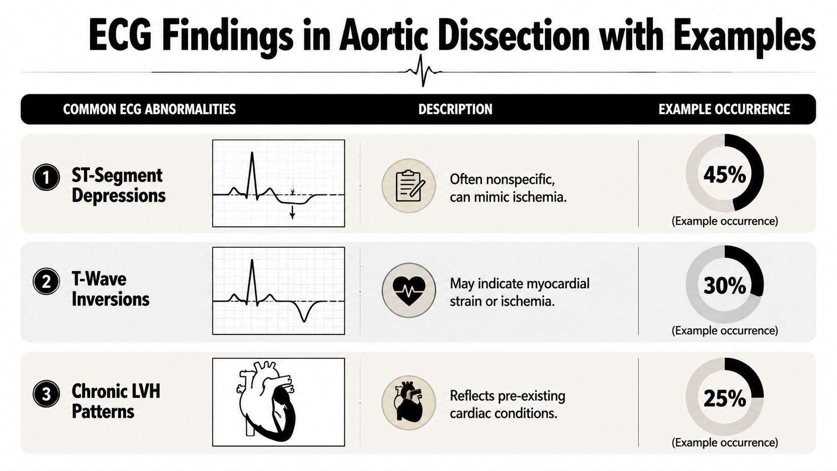
The ECG patterns in aortic dissection are often messy rather than dramatic. In one clinical review, ECG abnormalities were reported in about 70% of acute aortic dissection cases, but only 1% to 2% showed acute ST-elevation (Emergency Medicine Cases on ECG in aortic dissection). That means most traces are not clean textbook examples.
The most common patterns you may see
A wearable ECG may show ST-segment depression or T-wave inversion, which are nonspecific and can look like ischemia. A person might check their watch after sudden upper back pain and see a flattened or inverted repolarization pattern, then assume it must be “just stress” or “maybe nothing.” Those changes can't diagnose dissection, but they do tell you the tracing is not fully reassuring.
Some patients already have chronic left ventricular hypertrophy patterns, which can blur the picture further. If you've had high blood pressure or long-standing heart strain, the baseline ECG may already look abnormal, so a new change can be harder to recognize on a consumer device. A wearable can still be valuable here because comparison over time matters.
Conduction abnormalities can appear too. They're less famous than ST changes, but they add to the sense that the heart's electrical picture is not normal. The key is not to chase a single pattern. You're looking for unexpected change, especially when symptoms are severe.
A rare but important exception is true ST-elevation. It can happen, and that's where clinicians can get pulled toward an acute MI pathway too early. But it's uncommon enough that the ECG should be read with caution, not certainty.
For a deeper look at how one abnormal segment is tracked on consumer devices, see this guide to ST-segment monitoring with wearable ECGs. The point isn't to diagnose dissection from home. The point is to notice when a tracing looks meaningfully different from your usual baseline.
Limitations of ECG and Acute MI Mimics
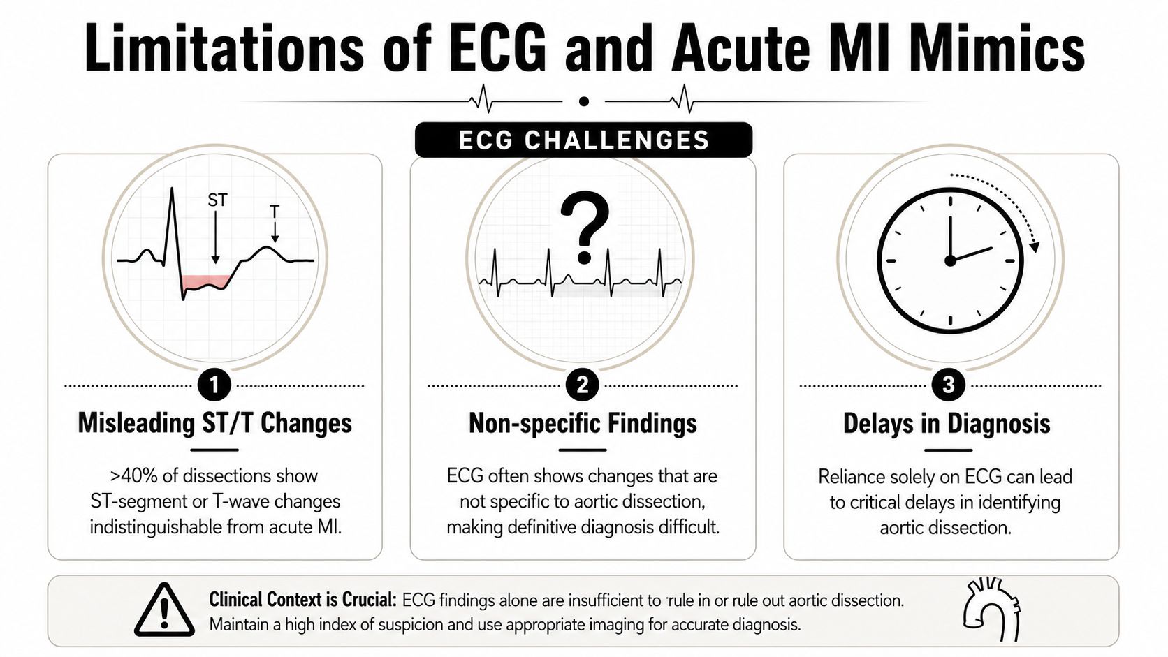
The main challenge is overlap. In one report, the 12-lead ECG was completely normal in only 30% of patients with dissection, while more than 40% had ST-segment or T-wave changes (Journal of Cardiology PDF00061-4/pdf)). Those are the same kinds of changes that can pull attention toward acute coronary syndrome.
Why the mimic matters
Dissection can resemble a heart attack because the heart may be under strain, or because the coronary arteries are involved. The tracing can point in two directions at once, which is why ECG findings alone are not enough to settle the question. Some dissections even show patterns that look like acute myocardial infarction, so the risk goes both ways, false reassurance and false direction.
That overlap can delay the right imaging. If chest pain comes from a torn aorta, the next decisive test is cross-sectional imaging, not another look at the watch. The ECG helps raise concern, but it cannot show whether the vessel wall is intact.
ECG is best for risk stratification, not confirmation.
Wearable users often ask whether a “normal” tracing means it is safe to wait. It does not. A dissection can still be present when the ECG looks fine, so symptoms have to lead the decision.
A useful way to hold the difference in mind is this. A heart attack is often an electrical story first, then a blood-flow story. Aortic dissection is a blood-vessel story first, then sometimes an electrical story. The ECG can catch the echo, but not the torn wall itself.
Recognizing Urgent Signs and When to Seek Care
A smartwatch ECG should never be the only thing you trust if the symptoms are alarming. A sudden tearing chest or back pain, pain that spreads to the abdomen, fainting, or sudden neurologic changes should override any reassuring screen. If the pain is severe and abrupt, the safest move is urgent medical evaluation.
Red flags that should not wait
- Severe, sudden pain: Chest, back, or abdominal pain that arrives fast and feels different from usual muscle pain.
- Neurologic symptoms: Weakness, confusion, trouble speaking, or collapse.
- Circulation clues: A pulse difference between arms or a major blood pressure difference if one is measured.
- ECG mismatch: A normal wearable ECG when the body feels seriously wrong, or an ECG with new ischemia-like changes.
Normal ECGs occur in 11% to 16% of aortic dissection cases, and dissections account for about 0.1% of STEMI presentations when the right coronary artery is involved (LITFL on acute aortic dissection). So even when the tracing looks clean, dissection can still be there, and even when the ECG looks like a STEMI, the root cause may be different.
If you're sorting through symptoms on your own, this guide to cardiac event symptoms can help you compare what you feel with patterns that deserve urgent attention. Use it as a checkpoint, not as a way to talk yourself out of care.
Bottom line: severe sudden pain plus a suspicious body exam means emergency evaluation, even if the watch says “normal.”


Diagnostic Pathway Beyond ECG
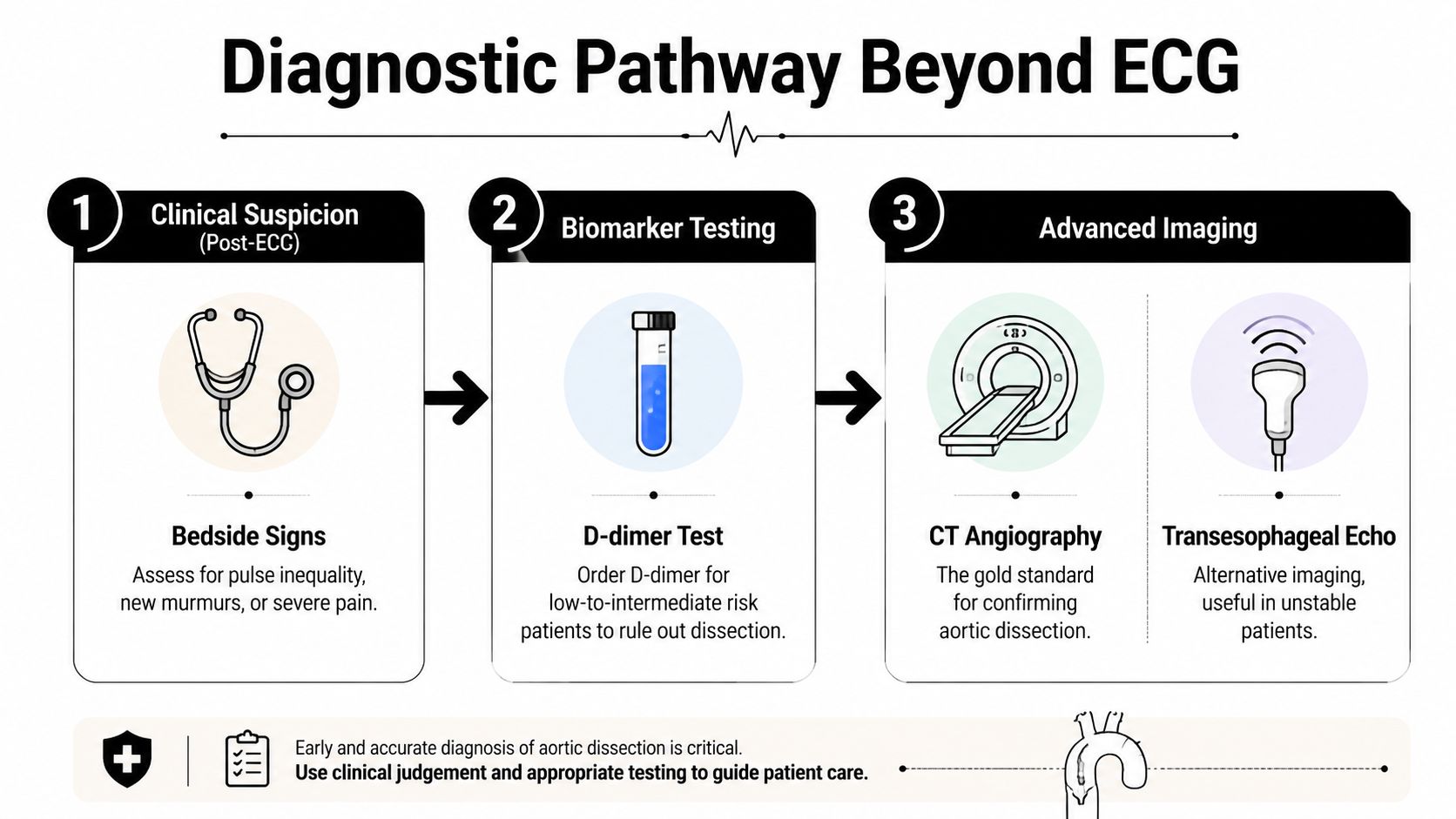
Once the ECG raises concern or fails to reassure, the next decision is clinical, not cosmetic. The diagnosis moves from “could this be dissection?” to “how likely is it, and what test will answer it safely?” That's where bedside findings, biomarkers, and imaging each do a different job.
From suspicion to imaging
First comes clinical suspicion. Pulse inequality, a new murmur, shock, or severe migrating pain all raise the stakes. These findings don't prove dissection, but they tell the team that the ECG is only one piece of a more dangerous puzzle.
Next comes biomarker testing in the right risk group. The ADvISED study evaluated 1,850 patients with suspected acute aortic syndromes and used the Aortic Dissection Detection Risk Score with D-dimer <500 ng/mL as a negative threshold. In patients with ADD-RS=0 and negative D-dimer, failure was 0.3%, and with ADD-RS<1 plus negative D-dimer, failure was also 0.3% (ADvISED study summary). That makes D-dimer a useful rule-out aid in selected low-risk situations, not a universal answer.
Then comes advanced imaging. CT angiography is the usual gold standard for confirmation, because it gives a direct view of the aorta and can show the tear and its extent. If a patient is too unstable for CT, transesophageal echocardiography can help at the bedside. MRI can also characterize the aorta, but it's not the first choice when speed matters.
The pathway works because each step narrows uncertainty. ECG helps identify danger, D-dimer can reduce unnecessary imaging in carefully selected low-risk patients, and CTA or TEE closes the diagnostic loop when the suspicion stays high.
If you want a practical overview of how CT is used in acute cardiac evaluation, this cardiac CT scan guide is a useful companion. For dissection, though, the key is urgency. A delayed image is a missed opportunity.
Practical Guidance for Wearable ECG Users
Your wearable is best used as a documentation tool, not a diagnosis machine. If you capture a tracing during chest pain, back pain, fainting, or a sudden “something is very wrong” moment, save it with the symptom time and what you were doing. That context often matters as much as the waveform.
What to flag
- New ST or T changes: Especially if they look different from your usual recordings.
- Unexpected conduction delay: A new sluggish-looking tracing, or a rhythm that feels off in a way you haven't seen before.
- Sudden rhythm instability: Fast, slow, or irregular patterns that appear with pain, weakness, or shortness of breath.
If the symptoms are severe, don't wait for an interpretation report before seeking emergency care. If the situation is less dramatic but still concerning, sharing the tracing with a human reviewer can help you organize what happened before your next visit.
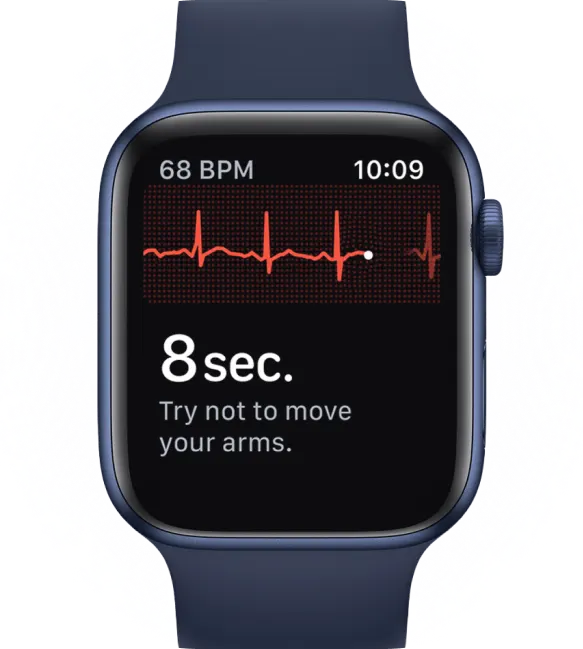
Use this Apple Watch ECG capture guide if you need a cleaner recording during a non-emergency moment. A better tracing won't rule out dissection, but it can make the next conversation with a clinician much more useful.
The main goal is simple. Keep the ECG as evidence, not as reassurance when the body is sending an alarm. If you're in doubt, treat the symptom pattern seriously first.
Conclusion and Next Steps
Aortic dissection is one of those conditions where the ECG can help, but it can also mislead. A normal tracing does not exclude it, abnormal ST or T changes can mimic a heart attack, and a definitive diagnosis often comes from urgent imaging rather than more guessing. That's why the safest mindset is calm, fast, and symptom-driven.
For wearable users, the best habit is to pair the ECG with context. Save the tracing, note the symptom, and pay attention to severe sudden pain, neurologic changes, or circulation differences between arms. If those show up, the watch is not the decision maker, emergency care is.
If you want help making sense of wearable tracings after the immediate danger has passed, upload them to Qaly for human-reviewed analysis and interval tracking, then bring those reports to your clinician. It's a strong way to bridge home monitoring with professional judgment, especially when your symptoms don't fit neatly into a simple box.
When your body sends a warning, trust the warning first, then use the data to guide the next step.

Unsure about an ECG? Certified experts review it within minutes, day or night.



.png)
.png)