


Key Takeaways

User Profile
If you're reading this with an Apple Watch, Kardia device, Fitbit, Samsung watch, or another wearable nearby, your question may be very simple and very human: if something serious happened to my heart, would my device catch it?
That concern makes sense. Medical language can feel cold, and terms like pulseless electrical activity can sound terrifying until someone slows down and translates them into plain English.
What Is Pulseless Electrical Activity
Hello Heart Hero. Pulseless electrical activity, often shortened to PEA, means the heart still shows electrical activity, but it isn't producing an effective pulse.
A simple way to picture it is a light switch and a light bulb. The switch is on, so electricity is flowing. But the bulb is broken, so the room stays dark. In PEA, the heart's electrical system is sending signals, but the heart muscle isn't creating enough pumping action to move blood through the body.
Blood flow is what keeps the brain, heart, and other organs alive. Electrical activity by itself isn't enough. The body needs the heart to squeeze effectively.
According to Cleveland Clinic's overview of pulseless electrical activity, PEA accounts for about 20% of sudden heart-related deaths outside hospitals and is the initial rhythm seen in 40 to 60% of in-hospital cardiac arrests. That tells us PEA isn't rare, especially in hospitals where teams are watching closely enough to identify it.
Why the name sounds confusing
Many people hear the word "electrical" and assume the heart must still be working. That's the trap. Electrical activity and blood flow are not the same thing.
A heart monitor can show an organized rhythm. A wearable ECG can sometimes record electrical patterns too. But if the heart muscle doesn't convert that signal into a meaningful squeeze, there may be no pulse.
Practical rule: In PEA, the monitor may look active while the body is in a life-threatening emergency.
Why people studying wearable ECGs care about this
If you use a watch or portable ECG, learning the causes of pulseless electrical activity helps you set the right expectations. These tools can be useful for rhythm awareness. They are not a substitute for checking circulation, symptoms, or getting emergency help when someone collapses.
That distinction can lower anxiety in a helpful way. It gives you a clearer sense of what your device can do well and where its limits begin.
When the Spark Is There But the Engine Is Not
Your heart has two jobs happening together. First, it creates an electrical signal. Second, the heart muscle responds to that signal by pumping blood.
A car analogy helps. The spark plug fires, and that spark helps the engine move. If the spark is present but the engine is seized, the car won't go anywhere. PEA is that kind of disconnect.
The heart's wiring and the heart's squeeze
The electrical part starts with the heart's natural pacemaker and conduction system. That signal travels through heart tissue in an orderly way. On an ECG, that can look organized.
The mechanical part is the actual squeeze of the heart muscle. That squeeze creates blood pressure and a pulse you can feel at the wrist or neck.
In PEA, those two parts are uncoupled. The wiring appears to be doing something. The pump is failing.
How PEA differs from other arrest rhythms
Many readers get mixed up here, especially if they've looked at ECG examples online.
- PEA: electrical rhythm is present, but there's no effective pulse.
- Asystole: there's no meaningful electrical activity and no pulse.
- Ventricular fibrillation: the electrical activity is chaotic, so the heart can't pump in a coordinated way.
These are all medical emergencies, but they are not the same problem. PEA is especially important because it often points to an underlying cause that may be reversible if a team identifies it fast enough.
Why an ECG alone can mislead you
An ECG answers one question well: what electrical pattern is present?
It doesn't directly answer another question: is the body being perfused with blood right now?
That gap is why someone can have an organized rhythm on a monitor and still be in cardiac arrest. Clinicians sometimes call this an electrical-mechanical mismatch. You don't need to memorize the phrase. The key idea is simpler than the jargon.
The heart can look "on" electrically while failing "off" mechanically.
That single idea explains why the causes of pulseless electrical activity matter so much. The rhythm itself isn't the whole story. The body is sending a distress signal that something deeper is preventing the pump from doing its job.
Finding the Why Behind PEA The Hs and Ts
PEA is often not the root problem. It's the end result of another crisis that has stopped the heart from pumping effectively. That's why emergency teams use a checklist called the 5 Hs and 5 Ts.
This framework helps clinicians move quickly through the most common reversible causes of pulseless electrical activity instead of guessing. The standard list includes hypovolemia, hypoxia, hydrogen ion acidosis, hypo or hyperkalemia, hypothermia, tension pneumothorax, tamponade, toxins, thrombosis, and trauma, as described in this ACLS review of PEA causes. The same review notes that respiratory problems leading to hypoxia accompany up to 50% of PEA cases.
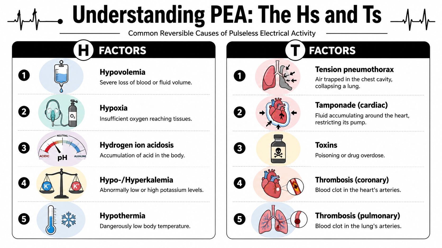
The H causes
Here is the first half of the checklist in plain language.
- Hypovolemia: The body doesn't have enough circulating fluid or blood. Severe bleeding is the classic example. If the heart has too little volume to pump, electrical activity may continue briefly while circulation fails.
- Hypoxia: The body isn't getting enough oxygen. This is one of the most important causes because the heart muscle and brain both depend on oxygen every second.
- Hydrogen ion acidosis: The blood becomes too acidic. When this happens, heart muscle and other tissues don't function normally.
- Hypo or hyperkalemia: Potassium is too low or too high. Potassium strongly affects how heart cells fire and recover. If you've ever wondered how electrolytes change ECG patterns, this guide on electrolyte imbalance and your ECG is a useful companion.
- Hypothermia: The body temperature drops dangerously low. Cold slows many body systems, including the heart.
A quick visual can help if you're a learner who remembers by seeing the pattern in action.
The T causes
The second half of the list includes structural or external problems that can physically block circulation.
- Tension pneumothorax
Air becomes trapped in the chest and compresses structures inside it. That pressure can interfere with blood returning to the heart. - Tamponade
Fluid builds up around the heart and squeezes it from the outside. The heart may receive electrical signals normally but still can't fill and pump well. - Toxins
Certain drugs, overdoses, or poisons can suppress the heart or circulation. - Thrombosis
This means a blood clot. The clot may be in the lungs or in the coronary circulation of the heart. - Trauma
A major injury can trigger several of these problems at once, including bleeding, chest injury, or obstructed circulation.
A bedside way to think about the list
You don't need to memorize every term like you're taking an exam. A simpler way to group the causes is this:
- Not enough blood or oxygen
- Chemistry is severely off
- Pressure is stopping the heart from filling or pumping
- A clot, toxin, or injury has disrupted circulation
Key insight: PEA often means "find the hidden problem" rather than "shock the rhythm."
That framing is important. It turns a frightening phrase into a logic problem. Clinicians ask what is starving the heart of oxygen, volume, space, or normal chemistry. Then they act on that cause.
Your Wearable ECG and the Limits of At Home Monitoring
This is the question many wearable users are really asking. Can my watch see PEA?
The honest answer is no. A wearable ECG records electrical activity. PEA is diagnosed when there is electrical activity and no pulse. The missing piece is the pulse.
Why the watch can't make the diagnosis
A watch can capture rhythm strips. Some devices can alert for certain rhythm patterns. That can be helpful in the right setting.
But pulseless electrical activity is not defined by the tracing alone. It's defined by a mismatch between what the monitor shows and what the body is doing. If there is no blood flow, a person is in crisis even if the rhythm strip still looks organized.


Where false reassurance can happen
A reader might think, "If my ECG doesn't look terrible, I must be okay." That's not a safe assumption in a collapse emergency.
If someone is unresponsive, not breathing normally, or has no pulse, the situation is urgent regardless of what a wearable screen suggests. Consumer devices are not built to replace emergency assessment.
For people who want a broader picture of what these devices can and can't do over time, this article on continuous monitoring with wearable heart tools is useful context.
The right role for a wearable
A wearable ECG can still be valuable. It can help document palpitations, irregular rhythms, or patterns that are worth discussing with a clinician.
It just can't answer the life-or-death bedside question that defines PEA: is the heart creating a pulse right now?
If a person collapses, symptoms and responsiveness matter more than the watch screen.
That's not a knock on wearable tech. It's using the right tool for the right job.
How Medical Teams Diagnose and Treat PEA
When a medical team suspects PEA, the response is structured. It may look fast from the outside, but there is a clear sequence behind it.
First, they check responsiveness, breathing, and pulse. If the person has no pulse, they begin CPR. At the same time, they look at the monitor. If they see an organized electrical rhythm without a pulse, that points toward PEA.
What happens next in real time
The team doesn't stop at naming the rhythm. They start searching for the cause.
One clinician may manage compressions. Another may secure the airway or support breathing. Another may review the situation for clues such as trauma, bleeding, overdose, breathing failure, or signs of a clot. In some cases, bedside ultrasound or a transthoracic echocardiogram can help show whether the heart is barely moving, compressed, or structurally obstructed.
Why the environment matters
This kind of care depends on preparation, equipment, and quick access to the patient. In emergency departments and acute care areas, the physical setup matters more than many people realize. Reliable access to resuscitation space, positioning, and transport support can make a practical difference, which is why facilities pay close attention to essentials such as emergency bed supplies from Labs USA.
The process is more logical than it looks
From the outside, emergency care can seem chaotic. Inside the room, people are often working from a disciplined mental checklist.
- Confirm the arrest: no pulse, no effective circulation
- Support circulation: CPR begins immediately
- Read the rhythm: organized activity without pulse suggests PEA
- Search for the cause: the Hs and Ts guide the next actions
- Reverse what can be reversed: oxygen, fluids, procedures, or targeted treatment depending on the problem
Good emergency care for PEA is less about guesswork and more about rapid problem-solving.
That doesn't mean every case ends well. It does mean there is a framework behind the response, and that framework exists because the causes of pulseless electrical activity are often more important than the rhythm label itself.
What This Means for Your Heart Health Journey
Learning that a wearable can't diagnose PEA may feel disappointing at first. But it can, in fact, make your heart tracking more useful, because it sharpens your expectations.
A wearable isn't a home code cart. It's a personal monitoring tool. Its strength is helping you notice patterns before they become part of a larger problem.
Where your device does help
If you get palpitations, skipped beats, racing episodes, or strange interval changes, repeated recordings can give useful context. Trends matter. Timing matters. A saved rhythm during symptoms is often more informative than a vague memory at a later appointment.

A proactive approach also includes watching the bigger picture of cardiovascular risk. If you're trying to understand how long-term factors fit into day-to-day monitoring, this article on a risk score for cardiovascular disease can help connect those dots.
When to use your data and when to seek urgent care
Use your wearable data for questions like these:
- Symptom tracking: "What was my rhythm when I felt that flutter?"
- Pattern spotting: "Does this happen after exercise, stress, poor sleep, or a medication change?"
- Clinical conversations: "Can I bring this recording to my doctor visit?"
Do not rely on it alone when symptoms are severe.
- Call emergency services: if someone collapses, is unresponsive, has severe chest pain, or isn't breathing normally
- Get urgent medical help: for fainting, sudden major shortness of breath, or alarming new symptoms
- Use the device as supporting information: not as a final verdict
Your watch can support awareness. It can't replace emergency judgment.
Empowered Knowledge for a Healthier Heart
PEA sounds technical, but the core idea is simple. The heart can show electrical activity and still fail to pump blood. That is why the causes of pulseless electrical activity matter so much.
Those causes are often organized into the Hs and Ts, a practical checklist that helps clinicians look for reversible problems such as low oxygen, low volume, severe chemistry imbalance, pressure around the heart, toxins, clots, or trauma. For wearable users, the biggest takeaway is just as important. A home ECG can record electricity, but it can't diagnose "pulseless."
That knowledge isn't meant to scare you. It's meant to give you a cleaner mental model. When you understand what your device can do, and what it can't, you're in a better position to use it wisely, ask better questions, and act quickly when symptoms are serious.
Staying curious about your heart is a strength. Clear information beats false reassurance every time.
Using an Apple Watch, Kardia, Fitbit, or Samsung watch? Qaly reviews your ECG recordings and helps you track rhythm and interval changes over time


.png)
.png)