


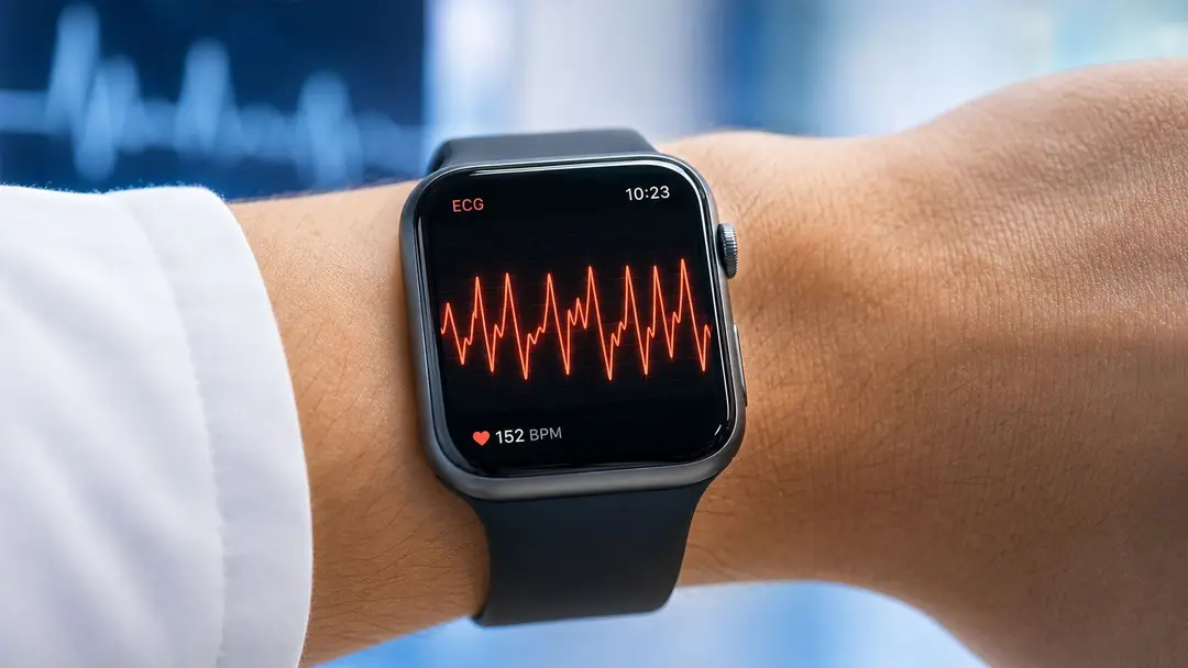


Hello Heart Hero. You glance at your watch, see a fast, irregular rhythm, and then notice the QRS shapes seem to keep changing. That's the kind of moment that can make your stomach drop, especially if you're already tracking palpitations, recovering from a heart event, or checking a new medication's effect on your rhythm.
If that's where you are right now, take a breath. A single-lead ECG can show a lot, but it can also leave out the context that decides whether a rhythm is dangerous, temporary, or just hard to classify. For people using Apple Watch, Fitbit, Kardia, Samsung, Withings, and similar devices, clarity matters more than guesswork. If you've also been reading about everyday palpitations and want a gentle overview of common causes, causes and relief for palpitations is a useful companion read.
One reason this gets confusing is that a wearable can show a wide-complex rhythm without telling you why it happened. If you've ever wondered whether your watch ECG is even seeing the right thing, this overview of watch ECG detection for ventricular tachycardia helps frame the limits and the value of a clean recording.
A Racing Heartbeat on Your Watch and What It Might Mean
You may have been sitting at your kitchen table, scrolling through a notification, or recovering after a workout when your watch flashed a tracing that looked messy and fast. Maybe the line wasn't just quick. Maybe the QRS complexes looked wide, uneven, and different from beat to beat. That's enough to make anyone stop and worry, and that reaction makes sense.
Hello Heart Hero again, and welcome. You're not overreacting by wanting a straight answer, especially if you've had palpitations before or you're trying to make sense of a monitor after a medication change. The goal here is simple, to help you read the pattern without getting lost in alarm.
This topic matters most for people using single-lead wearable ECGs, people checking rhythms after a cardiac event, and people who've been told to watch for side effects from heart-related drugs. Wearables can capture real information, but they can't replace clinical context. That matters a lot when the rhythm is wide and irregular.
A good way to think about the next few sections is this, a tracing can look frightening for several different reasons, and those reasons don't all mean the same thing. If you're anxious, that doesn't mean you're fragile. It means you're paying attention.
Practical rule: A watch ECG can be a strong clue, but it's not the final diagnosis when the rhythm is wide, fast, and changing shape.
What Polymorphic Ventricular Tachycardia Actually Is
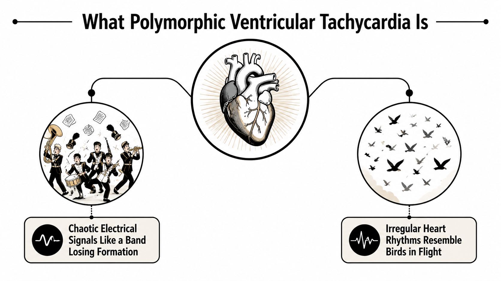
A normal rhythm has a clear pattern. Each beat arrives in a steady sequence, like a band keeping time together. In polymorphic ventricular tachycardia, that pattern falls apart, and the shape of the QRS complexes changes from beat to beat.
The changing shape is the key clue. A flock of birds can stay together while shifting direction in the sky, and the tracing behaves in a similar way, one fast rhythm, but with a moving outline. That is what polymorphic means here, the QRS pattern changes beat to beat.
How it differs from monomorphic VT
In monomorphic ventricular tachycardia, the QRS complexes look similar from one beat to the next. The tracing is repetitive, almost as if the same stamp were pressed again and again. In polymorphic VT, the shape does not stay fixed. A medical reference describes it as a rapid ventricular tachycardia with variable morphology and a continuously varying QRS pattern in a given lead (ScienceDirect medical reference).
That difference is not just visual. It tells clinicians that the lower chambers of the heart are firing in an unstable way. A major cardiology source describes polymorphic ventricular tachycardia as a malignant ventricular tachyarrhythmia because it can stop on its own or turn into ventricular fibrillation, which causes cardiac arrest. If arrest happens, treatment requires emergency defibrillation (AHA journal source).
Where torsades de pointes fits
You will often hear torsades de pointes mentioned with polymorphic VT. It is one form of polymorphic VT, usually linked to a long QT interval. The name refers to the twisting appearance of the QRS axis, but the practical point matters more than the label. The rhythm is unstable, and the cause shapes the treatment.
The difference also matters for people trying to make sense of a watch tracing. A single-lead wearable ECG can show a fast, irregular, changing rhythm, but it cannot reliably tell you whether the QT interval is prolonged. If you want a plain-language refresher on rhythm terms, see this guide to a heart rhythm disorder on Qaly.
The same visual pattern on a watch can come from different clinical problems, so the context matters as much as the tracing itself.
If you remember one sentence, make it this. Polymorphic VT is a fast ventricular rhythm with changing QRS shape, and that changing shape is what separates it from monomorphic VT.
The Two Different Faces of Polymorphic VT
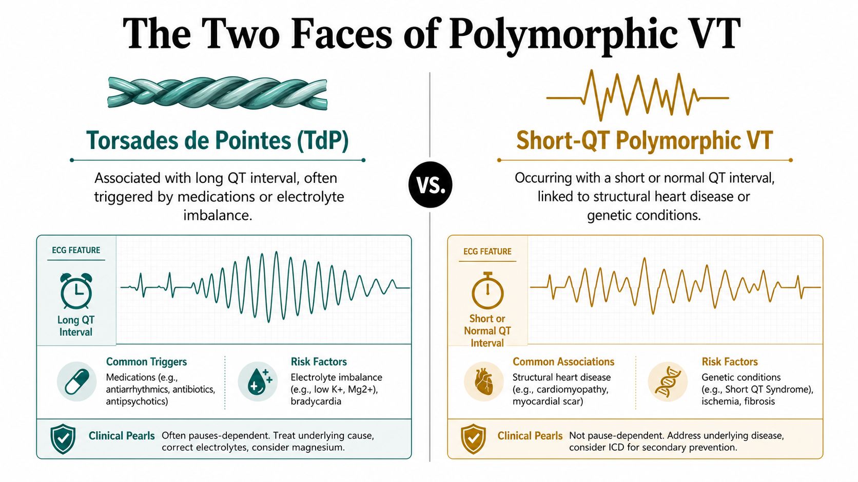
The term "polymorphic VT" is often misunderstood as a single rhythm. Clinically, it's better understood as two different entities. One has prolonged QT, often called torsades de pointes. The other happens without QT prolongation. They don't behave the same way, and they don't point to the same causes.
Why the QT interval changes the story
The QT distinction matters because it changes the trigger list. In QT-prolonged PMVT, the problem is usually tied to repolarization issues, often from medications or electrolyte problems. In non-QT-prolonged PMVT, clinicians think more about acute ischemia or unstable structural heart disease (European cardiology reference). That split is one reason single-lead wearables can be helpful but incomplete.
A watch tracing can show the rhythm, but it usually can't tell you whether the QT is long enough to point toward torsades. That's one of the biggest blind spots in consumer ECG interpretation. A single lead may reveal a wide, irregular rhythm, but it can't reliably tell you the underlying cause on its own.
Why this matters for at-home ECG users
If you're checking a wearable strip, the phrase “polymorphic VT” shouldn't end the conversation. It should start a question. Was the rhythm long-QT related, or was it something else? Did the person just start a new drug, lose electrolytes, or develop ischemia? Or does the pattern suggest something inherited?
For readers trying to understand QT issues more thoroughly, this guide to QT prolongation causes provides useful background without assuming a hospital setting.
The main point is simple. PMVT is not one single rhythm. It's a family of rhythms, and the QT interval helps separate two very different clinical paths.
Common Causes and Triggers Worth Knowing
A racing rhythm on a watch strip often starts with a clear trigger. A new prescription, a recent illness, a harder-than-usual workout, or a child who faints on the field can all sit behind the same broad label of polymorphic VT. The tracing may look similar, but the cause is often a different story.
Medication and electrolyte changes
Some episodes follow QT-prolonging medications or a shift in electrolytes, especially when potassium or magnesium runs low. That is why a new antibiotic, antiarrhythmic, antidepressant, or nausea medicine deserves attention if palpitations start soon afterward. If you want a practical overview of magnesium-related clues, VitzAi.com signs of low magnesium can help you think through common symptoms without jumping to conclusions.
Medication problems do not always announce themselves loudly. Sometimes the first clue is a strange, fast rhythm on a wearable, especially if the QT interval is already vulnerable. A related pitfall is the R-on-T pattern, where an early beat lands on the vulnerable part of repolarization, a setup discussed in this Qaly explanation of R-on-T on ECG. That is why clinicians usually start with a careful medication review when they evaluate PMVT.
Ischemia, structural disease, and stress-triggered inherited risk
Non-QT-prolonged PMVT tends to raise concern for acute ischemia or structural heart disease. That can fit someone with chest pressure, a recent coronary event, or a known cardiomyopathy. A wearable may show the rhythm, but it will not explain whether the heart muscle is under strain from a blood flow problem.
Then there is catecholaminergic polymorphic ventricular tachycardia, or CPVT. It is a rare inherited arrhythmia syndrome, estimated to affect about 1 in 10,000 people worldwide (MedlinePlus Genetics). Episodes often begin in childhood and are triggered by exercise or emotional stress. In untreated or high-risk cohorts, reviews report mortality rates of 30% to 50% by age 35 (AER review).
A child who faints during sports, a teen who gets palpitations during competition, or an adult with a normal resting tracing but collapse during exertion all fit a pattern that deserves careful attention. For CPVT, the resting ECG is often normal, so a wearable taken at rest can miss the problem completely. Genetic testing has an approximate 60% yield, and RYR2 and CASQ2 account for most known cases (Merck Manual).
A normal resting wearable ECG does not rule out CPVT if the symptoms only show up during exercise or emotional stress.
For a watch user, that is the red-flag pattern to remember.
Symptoms, Red Flags, and What It Looks Like on a Wearable ECG
A person with polymorphic VT may notice palpitations, dizziness, breathlessness, chest pressure, or a sudden feeling that something is wrong. Sometimes the first clear event is syncope, which means fainting. In CPVT, these symptoms often appear during exercise or strong emotion, not while resting.
A wearable ECG can capture a fast wide-complex rhythm that changes shape from beat to beat. To a non-expert, it may look irregular and chaotic, with the width and outline of the QRS complexes shifting as the rhythm speeds along. That changing pattern is what clinicians mean by variable morphology and a continuously varying QRS pattern in a given lead.
The part a watch cannot provide is the full clinical context. A single-lead tracing usually cannot tell torsades from another form of polymorphic VT on its own. It also cannot reliably show whether the cause is QT prolongation, ischemia, structural heart disease, or CPVT. The strip is only one piece of the story.
When to treat it as urgent
Emergency help is needed if the rhythm comes with fainting, collapse during exercise, severe chest pain, or palpitations that stay rapid and do not settle. Those symptoms should not be brushed off. If someone becomes unresponsive, emergency defibrillation may be needed because polymorphic VT can turn into ventricular fibrillation and cardiac arrest.
If the symptoms were brief but the tracing still showed a fast wide-complex rhythm, it still deserves review. A clear recording is more useful than trying to rely on memory alone, especially when the episode felt sudden or frightening.
What it looks like on the strip
On a watch ECG, the pattern often looks messy, with each wide beat changing slightly from the one before it. That is different from a steadier wide rhythm where each complex keeps the same shape. One simple way to picture it is that the beat outline keeps shifting, while a more regular wide rhythm repeats the same outline over and over.
That visual pattern matters, but it should not be interpreted in isolation. The symptoms, the setting, and the timing help decide whether the tracing is pointing toward a QT-related episode, a stress-triggered inherited rhythm problem, or another cause. A normal resting wearable ECG can still miss CPVT if the episodes only happen with exercise or emotional stress.
Get your ECG checked by certified experts within minutes on the Qaly app.


How Doctors Diagnose and Manage Polymorphic VT
Doctors usually start with a 12-lead ECG, telemetry, and a careful look at the QTc. They also check electrolytes, review medications, and look for ischemia or structural heart disease with tests like echocardiography. If the story points toward CPVT, they may use exercise testing and genetic testing.
What happens in the urgent setting
If the rhythm is sustained or the person is unstable, treatment is immediate. That can include defibrillation for arrest, IV magnesium for torsades, stopping the offending drug, correcting electrolytes, and treating ischemia if that's the trigger. The exact treatment depends on the subtype, which is why the QT distinction matters so much.
What follows after the emergency
For CPVT, beta-blockers are the cornerstone of therapy, and flecainide is often added when symptoms continue. High-risk patients may also be considered for an ICD, depending on the full clinical picture. The point isn't to memorize every option. It's to understand that polymorphic VT is managed by identifying the trigger, not just suppressing the rhythm.
Wearable ECGs fit into this process in a practical way. A clean strip taken during symptoms can help clinicians see the timing, the rate, and whether the rhythm really is wide and variable. That can save time, especially when symptoms are brief or unpredictable.
One useful option for people using consumer devices is Qaly, which connects wearable ECG recordings to certified cardiographic technicians for human review and interval measurements like PR, QRS, and QTc. That kind of review doesn't replace medical care, but it can make the handoff to a clinician much clearer.
Practical Next Steps for At-Home ECG Users
If your watch or handheld ECG captures a fast wide-complex rhythm, record it again if you can do so safely. Sit still, keep the device steady, and try to capture the clearest strip possible. A shaky recording is still worth keeping, but a cleaner one makes rhythm review much easier.
What to do tonight
- Save the tracing immediately. Don't delete a strange strip just because it scared you.
- Write down what was happening. Note exercise, stress, a new medication, fainting, chest pain, or breathlessness.
- Check whether symptoms are ongoing. If there's collapse, severe chest pain, or fainting, seek emergency care now.
- Share the recording with a clinician or certified reviewer. A technician can often help sort out whether the strip looks like a wide-complex tachycardia, whether the QRS is changing beat to beat, and whether QTc looks concerning.
- Track the pattern over time. Recurrent episodes during exertion, especially with a normal resting ECG, deserve more attention than an isolated resting alert.
When CPVT deserves extra urgency
If the story includes childhood fainting during exercise, a family history of sudden cardiac death, or a normal resting ECG with exertional symptoms, don't brush it off. That pattern fits CPVT far more than an ordinary skipped-beat problem. Even a reassuring resting tracing can miss it completely.
If you want a bridge between your wearable and your clinician, a human-reviewed ECG service can help. It can give you a readable report, interval measurements, and a record you can bring to your next appointment. That can turn a scary notification into something your care team can work with.
If your watch ECG has shown a fast wide rhythm, don't sit with uncertainty for days. Capture the cleanest tracing you can, note what you were doing, and arrange a same-day or next-day review if you're stable. If the episode included fainting, collapse, or chest pain, seek emergency care now.
Unsure about an ECG? Certified experts review it within minutes, day or night.


