


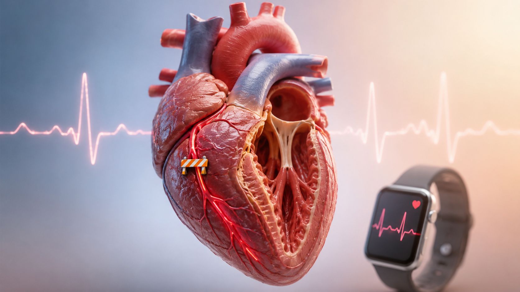


Key Takeaways

User Profile
Hello Heart Hero. If you've just noticed a strange flutter on your watch, or you glanced at a single-lead ECG and thought, “That doesn't look normal, but I can't tell what it means,” you're exactly the kind of reader this is for. A lot of people are trying to make sense of a possible occlusion of the right coronary artery while feeling tired, uneasy, or frankly skeptical about whether a rushed appointment will give them a real answer.
You're not overreacting for wanting clarity. The right coronary artery can matter in ways that don't always look dramatic at first, and wearable ECGs can catch useful clues without telling the whole story. The trick is knowing what those clues mean, what they can't prove, and when a symptom deserves urgent care instead of more guessing.
This guide walks step by step from the anatomy of the artery, to the ECG patterns doctors look for, to the symptoms people often miss, and finally to how a wearable ECG fits into real-life decision-making. The goal is simple, help you feel informed, not panicked.
A Warm Hello From Your Heart Guide
A person can be sitting at home, feeling a little off, and see a strange rhythm on a watch ECG that looks important but not obviously dangerous. Maybe it was a flutter after climbing stairs. Maybe it was a burst of nausea, a wave of lightheadedness, or that “something isn't right” feeling people often struggle to describe.
Hello Heart Hero. If that sounds familiar, you're in the right place. You don't need to know medical jargon to start understanding what the right coronary artery does, why a blockage there can show up in sneaky ways, and how a wearable ECG can help you gather information without trying to diagnose yourself on the spot.
The important part is this, skepticism is understandable. If you've ever felt brushed off, or told your symptoms were probably stress before anyone looked carefully, it makes sense to want a clearer path. Here, the focus is on plain language and practical steps, not judgment.
What we're really talking about
An occlusion of the right coronary artery means blood can't get through that vessel the way it should. In some people, that shows up as classic chest pressure. In others, the clue is a watch ECG captured during symptoms, or a combination of nausea, breathlessness, and a slow heart rate.
The sections below move from the basics of the artery itself, to the ECG patterns that raise concern, to what symptoms deserve attention, and then to what to do with the recording on your wrist. If you've ever wondered whether your device is “enough,” you'll get a clear answer, along with the parts it can and can't do.
Understanding the Right Coronary Artery
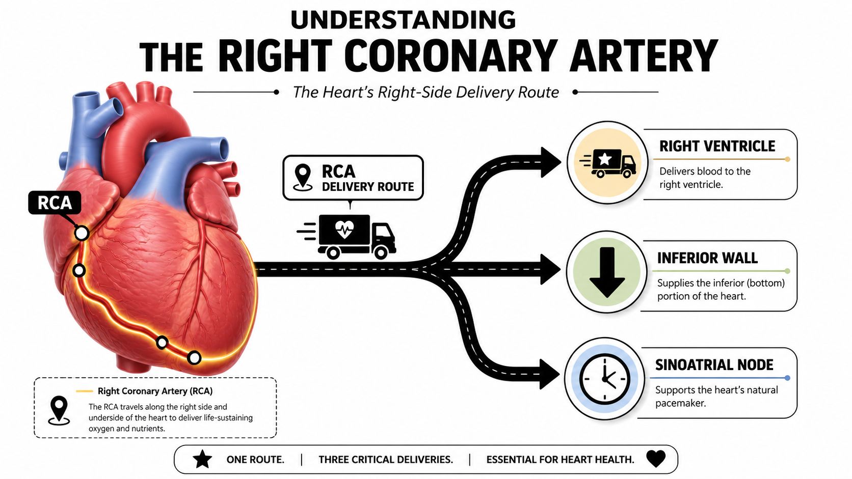
Think of the right coronary artery (RCA) as one of the heart's delivery routes. It runs along the right side and underside of the heart, sending blood to the right ventricle and, in many people, the inferior wall and back portion of the left ventricle. It also often supplies the sinoatrial node, which helps set the heart's rhythm.
When that route narrows or closes, the heart muscle downstream gets less oxygen-rich blood. That's what doctors mean by an occlusion, a blockage that stops blood flow. If the closure has been there for three or more months, it's called a chronic total occlusion (CTO), meaning the artery has been fully blocked for a long time rather than suddenly shut off Cleveland Clinic explains CTOs as complete blockages lasting three or more months.
A useful mental picture is a road closure versus a long-term detour. A sudden closure can create an emergency. A long-term blockage may let the body build collateral circulation, which is like side streets that partly reroute traffic. But those side streets often don't carry enough flow to fully supply the muscle, so people can still have ischemia and angina even when the artery looks “completely blocked” AHA review of RCA CTO physiology.
Dominant and nondominant patterns
People often find this confusing. The impact of an RCA blockage depends partly on whether the artery is dominant or nondominant. In a dominant pattern, the RCA supplies more of the bottom part of the heart. In a nondominant pattern, the territory is smaller, but that doesn't make it harmless.
Plain-language rule: a smaller artery can still cause a big problem if it affects the right ventricle or the heart's rhythm system.
That's why doctors pay attention to the artery's path, the symptoms, and the ECG together, not just one piece in isolation. For a broader overview of coronary disease, this coronary artery disease guide is a helpful companion read.
How an RCA Occlusion Shows on an ECG
The classic pattern doctors think about is inferior ST elevation, usually in leads II, III, and aVF. Those leads look at the bottom part of the heart, so when the RCA is involved, they can show injury there. Reciprocal changes, meaning opposite-looking shifts in other leads, can strengthen the suspicion that this is a real ischemic event rather than just noise.
One useful detail is that ST elevation in lead III greater than lead II can point toward an RCA culprit. That clue matters because the exact lead pattern can help separate one coronary territory from another. In some cases, bradyarrhythmias or right-sided involvement appear too, especially if the artery supplies the conduction system or the right ventricle multicenter RCA CTO registry insights.
What your watch can show, and what it can't
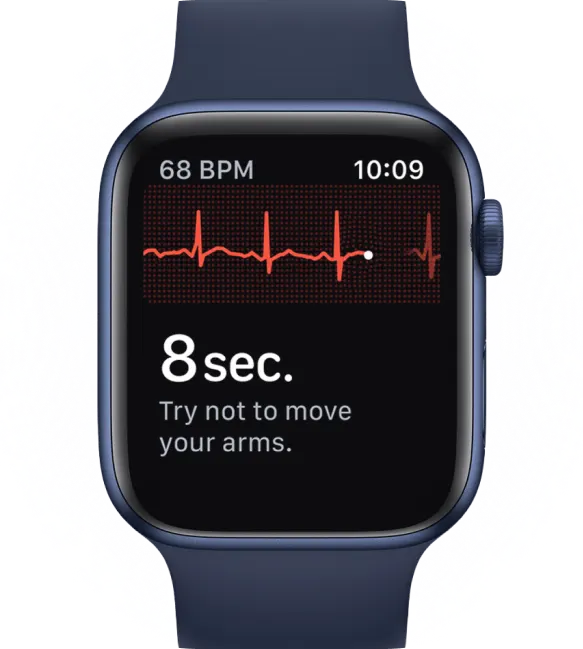
A wearable or single-lead ECG is useful for rhythm and rate, especially if symptoms come and go. It can capture a moment of palpitations, a pause, or a fast rhythm that might otherwise be gone by the time you reach care. But it can miss inferior changes, and a single lead can confuse an ischemic tracing with motion, poor contact, or plain noise.
That doesn't make your device pointless. It means you should use it as a recorder, not as a judge.
Capture a clean tracing during symptoms if you can, then stop trying to self-diagnose the squiggles in the moment.
For a practical primer on interpretation basics, this ECG reading guide can help you understand why one tracing may be informative while still needing clinical context.
Symptoms Worth Paying Attention To
A man comes in saying he thought it was indigestion. He felt nauseated, sweaty, and “just off,” then noticed his heart rate dropping on his watch. He didn't have the movie-version chest clutching, so he almost ignored it. That's how RCA problems can slip past people, especially when the symptoms are quieter than expected.
The classic warning signs still matter, chest pressure, pain that spreads to the jaw or arm, cold sweat, and breathlessness. But RCA events can also look more subtle. People may feel unexplained fatigue, lightheadedness, indigestion-like discomfort, or a vague sense that something is wrong.
Why the symptoms can look different
The RCA can involve the inferior wall and the heart's rhythm system. When that happens, a person may develop bradycardia, fainting, or other rhythm changes instead of the loud, crushing pain many people expect. A case-based review of nondominant RCA occlusion noted that isolated right ventricular infarction is uncommon, reported in only 1% to 3% of myocardial infarctions at autopsy and 5% to 15% of left ventricular infarctions Wiley review of nondominant RCA occlusion and ventricular tachyarrhythmias.
That rarity can be part of the problem. People, and sometimes clinicians, may underestimate an event because the symptoms don't match the stereotype. The heart doesn't care whether the presentation looks textbook.
A practical way to think about risk layering is this. New symptoms plus an abnormal wearable tracing plus a known history of coronary disease should be taken seriously, even if the discomfort is not dramatic. Not every symptom means an occlusion, but dismissing a pattern because it feels “atypical” can be dangerous.
If you've been brushed off before, keep records of what happened, what you felt, and how long it lasted. That history often matters more than a single snapshot.
What Happens in the Hospital and Why Time Matters
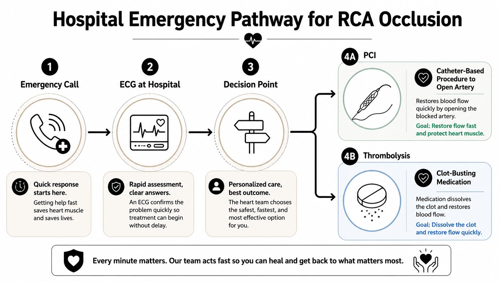
If the concern is an acute blockage, emergency teams usually move fast because opening blood flow sooner can limit damage. PCI, or percutaneous coronary intervention, uses a catheter to open the artery from inside. Thrombolysis uses clot-busting medicine to try to restore flow when PCI isn't immediately available or isn't the best option.
The choice depends on anatomy, timing, and the team available. A person who arrives early with an artery that can be reached in the cath lab may be sent for PCI. Someone whose situation or location makes PCI impractical may receive thrombolysis first. A chronic total occlusion is different, and in some people it's managed medically rather than reopened right away.
What the emergency flow usually feels like
The process is usually more organized than it feels from the outside. First comes the call for help, then an ECG, then a decision about whether the pattern and symptoms justify urgent reperfusion. The goal is to restore flow before more heart muscle is lost.
A quiet point that matters for worried readers, not every blocked RCA needs the same path. A chronic blockage, a delayed presentation, or anatomy that makes the procedure risky can change the plan. That isn't failure, it's the team choosing the safest option for that specific heart.
If you want a simple reference for how clinicians think about biomarker timing in chest pain workups, this troponin guide can help put the emergency evaluation into context.


Using Your Wearable ECG the Smart Way
The best time to record is during symptoms or right after they start, while you're still still and quiet. Sit down, rest your hands, and get a clean strip if your device allows it. Add a note about what you felt, such as chest pressure, nausea, dizziness, shortness of breath, or palpitations.
That small habit can turn a vague memory into useful clinical evidence. A watch strip by itself may not prove an RCA occlusion, but it can show rhythm changes, rate trends, or pauses that help a clinician decide what comes next. It can also help separate a real event from a brief sensation that passed quickly.
What human review adds
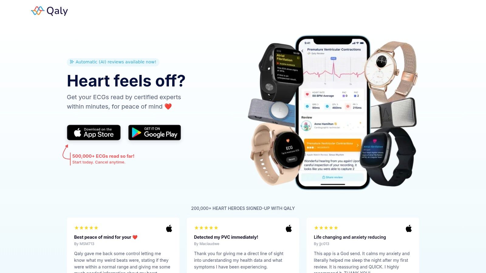
Consumer devices are good at collecting data, but they don't replace a clinician's eyes. Human-reviewed ECG services can turn a recording into a clearer narrative, often flagging rhythm issues such as atrial fibrillation, PVCs, or borderline intervals and giving you something organized to bring to an appointment. That's especially helpful if you've ever left a visit feeling like the conversation moved too fast.
Best use case: record the symptom, save the strip, and let a trained reviewer help translate the tracing into plain language.
Wearables still can't diagnose an occlusion on their own, and they can't replace a full 12-lead ECG when one is needed. They are tools for timing, documentation, and pattern recognition, not final diagnosis. If your symptoms are concerning, emergency services come first, not the app.
For readers who like to track changes over time, this ST segment monitoring guide gives useful context for what serial recordings can and can't tell you.
When You Feel Dismissed by the System
If you've had symptoms minimized before, that frustration is real. You shouldn't have to fight to be heard, and you also shouldn't have to walk into an appointment empty-handed and hope someone connects the dots for you. A short paper trail can make a real difference.
Keep a simple log with the date, what you felt, how long it lasted, and what your ECG showed. Bring a one-page summary that fits on a single page, not a stack of screenshots that nobody has time to sort through. Ask direct questions such as, “Could this be an inferior event given my tracing?” or “What should I do if this happens again?”
If you're also comparing devices, look for features that make recording easy and readable, and for some people, that includes options among the latest smartwatches from DigiDevice. The point isn't the brand itself, it's having a tool you can use quickly when symptoms start.
Pairing your own notes with a human-reviewed ECG report changes the conversation. Instead of “I feel funny,” you can say, “Here is what was captured, here is what was reviewed, and here is why I'm concerned.” That is calmer, clearer, and much harder to dismiss.
Your Take-Home Plan for the Next Time Something Feels Off
If you develop chest pressure with breathlessness, fainting, or sudden cold sweat with jaw or arm pain, call emergency services now. If palpitations come with dizziness and don't settle, that also deserves urgent attention. You don't need to prove it's a heart problem before asking for help.
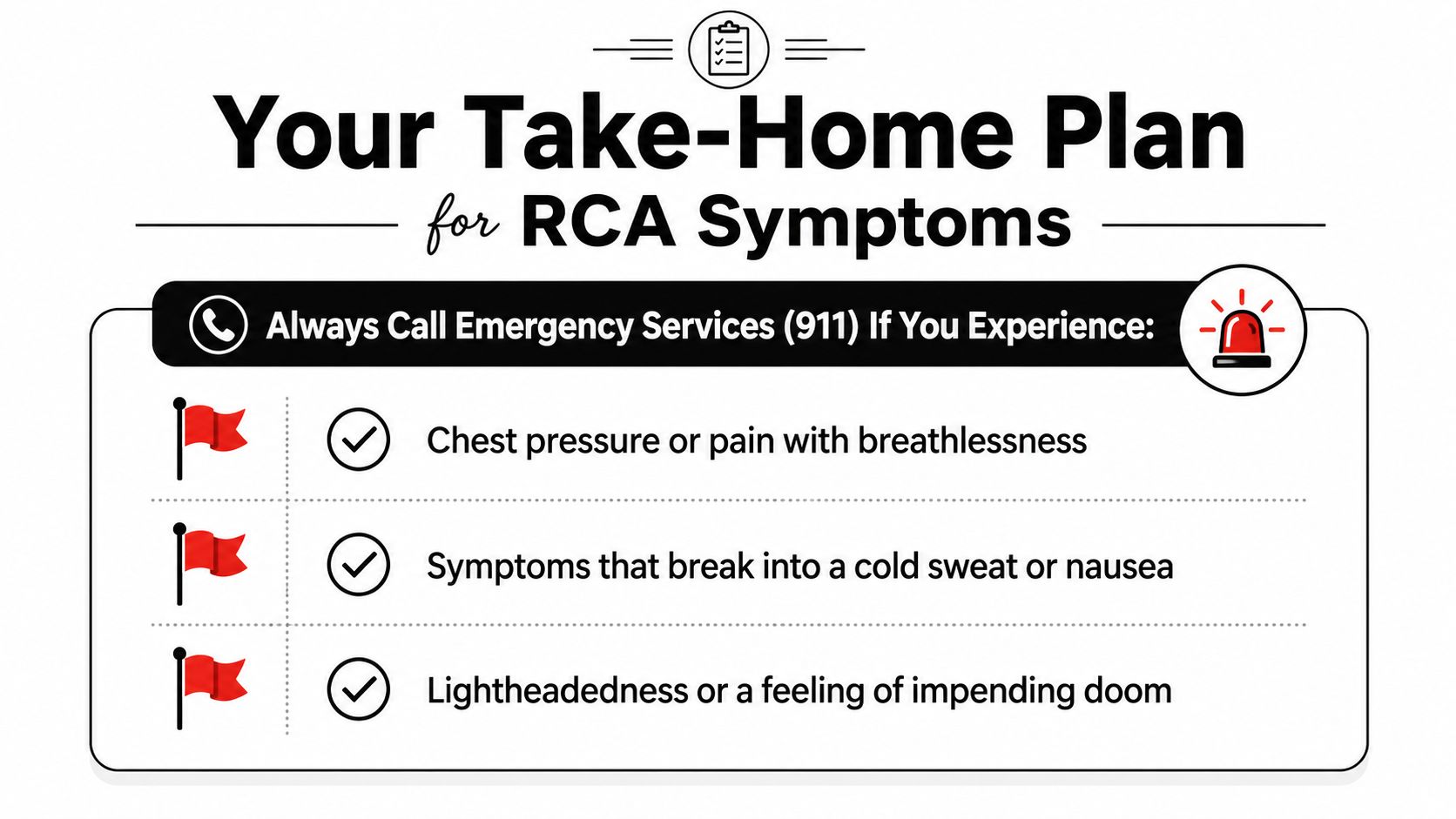
Three habits make wearable ECGs more useful:
- Record early: start the tracing while symptoms are active, not after they've faded.
- Keep a baseline: save a normal recording when you feel well so there's something to compare against later.
- Use human review: a clear report is easier to bring to a clinician than a raw strip that no one has time to interpret on the spot.
If you've been worried about the occlusion of right coronary artery, your caution isn't irrational. It's a sign that you're paying attention to your body, and that matters. Use your wearable as a careful witness, not a verdict, and if you want help organizing the ECG evidence before your next appointment, start with a service that offers human-reviewed readings so the conversation is grounded in something real.

Unsure about an ECG? Certified experts review it within minutes, day or night.



.png)
.png)